

Predicting language outcome and recovery after stroke: the PLORAS system
- PMID: 20212513
- PMCID: PMC3556582
- DOI: 10.1038/nrneurol.2010.15
Predicting language outcome and recovery after stroke: the PLORAS system
Abstract
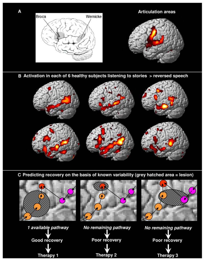
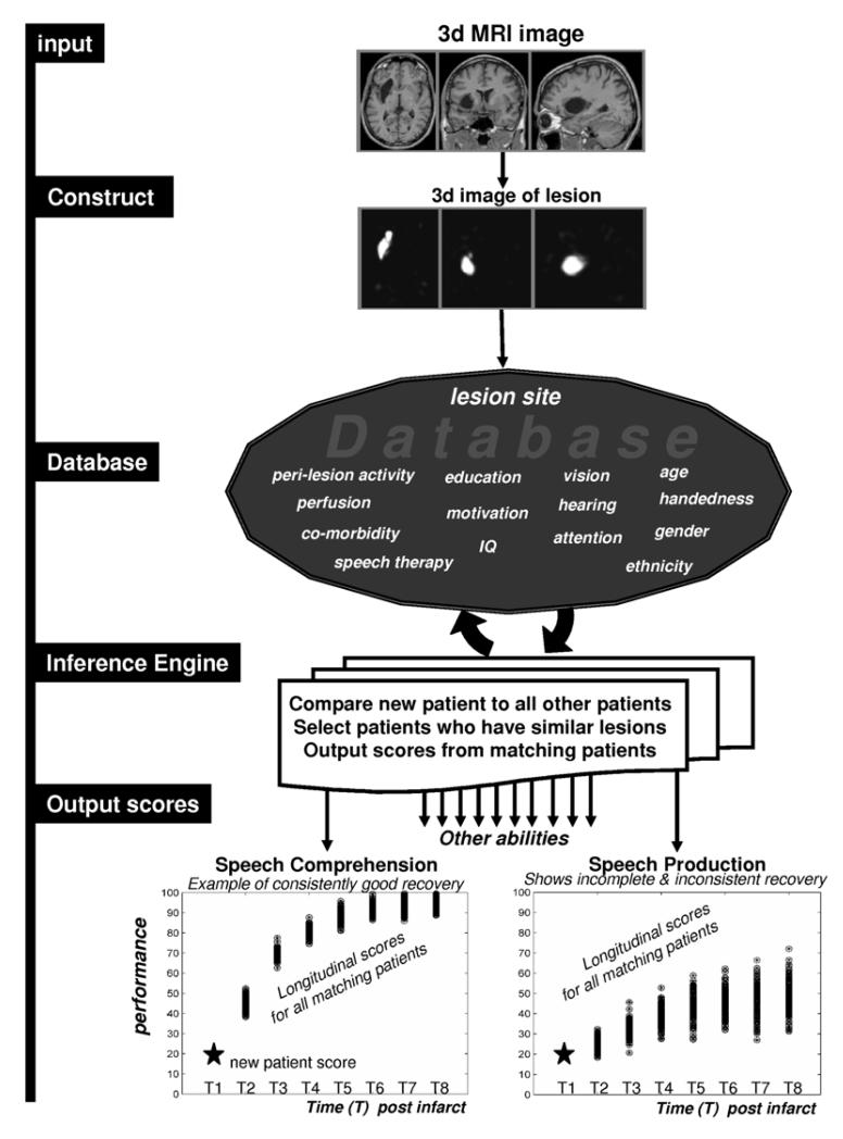
The ability to comprehend and produce speech after stroke depends on whether the areas of the brain that support language have been damaged. Here, we review two different ways to predict language outcome after stroke. The first depends on understanding the neural circuits that support language. This model-based approach is a challenging endeavor because language is a complex cognitive function that involves the interaction of many different brain areas. The second approach, by contrast, does not require an understanding of why a lesion impairs language; instead, predictions are made on the basis of the recovery of previous patients with the same lesion. This approach requires a database that records the speech and language capabilities of a large population of patients who have, collectively, incurred a comprehensive range of focal brain lesions. In addition, a system is required that converts an MRI scan from a new patient into a three-dimensional description of the lesion and compares this lesion against all others on the database. The outputs of this system are the longitudinal language outcomes of corresponding patients in the database. This approach will provide the patient with a range of probable recovery patterns over a variety of language measures.
Figures
Similar articles
-
The PLORAS Database: A data repository for Predicting Language Outcome and Recovery After Stroke.Neuroimage. 2016 Jan 1;124(Pt B):1208-1212. doi: 10.1016/j.neuroimage.2015.03.083. Epub 2015 Apr 14. Neuroimage. 2016. PMID: 25882753 Free PMC article.
-
Multivariate Connectome-Based Symptom Mapping in Post-Stroke Patients: Networks Supporting Language and Speech.J Neurosci. 2016 Jun 22;36(25):6668-79. doi: 10.1523/JNEUROSCI.4396-15.2016. J Neurosci. 2016. PMID: 27335399 Free PMC article.
-
Predicting language outcomes after stroke: Is structural disconnection a useful predictor?Neuroimage Clin. 2018 Mar 30;19:22-29. doi: 10.1016/j.nicl.2018.03.037. eCollection 2018. Neuroimage Clin. 2018. PMID: 30034998 Free PMC article.
-
Factors predicting post-stroke aphasia recovery.J Neurol Sci. 2015 May 15;352(1-2):12-8. doi: 10.1016/j.jns.2015.03.020. Epub 2015 Mar 20. J Neurol Sci. 2015. PMID: 25888529 Review.
-
Neurobiology of language recovery after stroke: lessons from neuroimaging studies.Arch Phys Med Rehabil. 2012 Jan;93(1 Suppl):S15-25. doi: 10.1016/j.apmr.2011.03.036. Arch Phys Med Rehabil. 2012. PMID: 22202187 Review.
Cited by
-
Recovery of Apraxia of Speech and Aphasia in Patients With Hand Motor Impairment After Stroke.Front Neurol. 2021 Mar 31;12:634065. doi: 10.3389/fneur.2021.634065. eCollection 2021. Front Neurol. 2021. PMID: 33868144 Free PMC article.
-
Clinical and neuroimaging factors associated with aphasia severity in stroke patients: diffusion tensor imaging study.Sci Rep. 2020 Jul 30;10(1):12874. doi: 10.1038/s41598-020-69741-1. Sci Rep. 2020. PMID: 32733102 Free PMC article.
-
Relative contributions of lesion location and lesion size to predictions of varied language deficits in post-stroke aphasia.Neuroimage Clin. 2018;20:1129-1138. doi: 10.1016/j.nicl.2018.10.017. Epub 2018 Oct 19. Neuroimage Clin. 2018. PMID: 30380520 Free PMC article.
-
Brain regions important for recovery after severe post-stroke upper limb paresis.J Neurol Neurosurg Psychiatry. 2017 Sep;88(9):737-743. doi: 10.1136/jnnp-2016-315030. Epub 2017 Jun 22. J Neurol Neurosurg Psychiatry. 2017. PMID: 28642286 Free PMC article.
-
Changes in auditory feedback connections determine the severity of speech processing deficits after stroke.J Neurosci. 2012 Mar 21;32(12):4260-70. doi: 10.1523/JNEUROSCI.4670-11.2012. J Neurosci. 2012. PMID: 22442088 Free PMC article.
References
-
- Sanai N, Mirzadeh Z, Berger MS. Functional outcome after language mapping for glioma resection. N Engl J Med. 2008;358:18–27. - PubMed
-
- Caplan D, et al. A study of syntactic processing in aphasia II: neurological aspects. Brain Lang. 2007;101:151–177. - PubMed
-
- Rijntjes M. Mechanisms of recovery in stroke patients with hemiparesis or aphasia: new insights, old questions and the meaning of therapies. Curr Opin Neurol. 2006;19:76–83. - PubMed
-
- Lazar RM, Antoniello D. Variability in recovery from aphasia. Curr Neurol NeurosciRep. 2008;8:497–502. - PubMed
Publication types
MeSH terms
Grants and funding
LinkOut - more resources
Full Text Sources
Medical
