. 2010 Dec;136(12):1891-900.
doi: 10.1007/s00432-010-0848-1. Epub 2010 Mar 6.

Long-term interferon-α treatment suppresses tumor growth but promotes metastasis capacity in hepatocellular carcinoma

Affiliations

Long-term interferon-α treatment suppresses tumor growth but promotes metastasis capacity in hepatocellular carcinoma

Peng-Yuan Zhuang et al. J Cancer Res Clin Oncol. 2010 Dec.

Abstract

Purpose: To elucidate the effect of IFN-α treatment on tumor growth and metastasis of hepatocellular carcinoma (HCC).

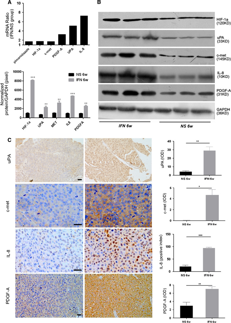
Methods: IFN-α administration was conducted in nude mice using an orthotopic implantation model of human HCC, and the key molecular markers in the IFN-α treatment was detected by immunohistochemistry staining and PCR array.

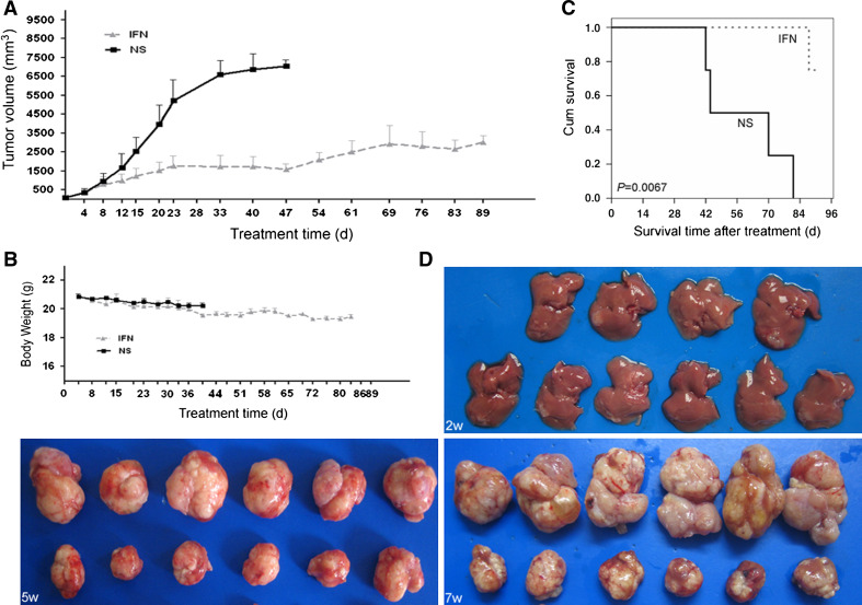
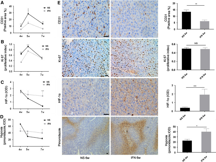
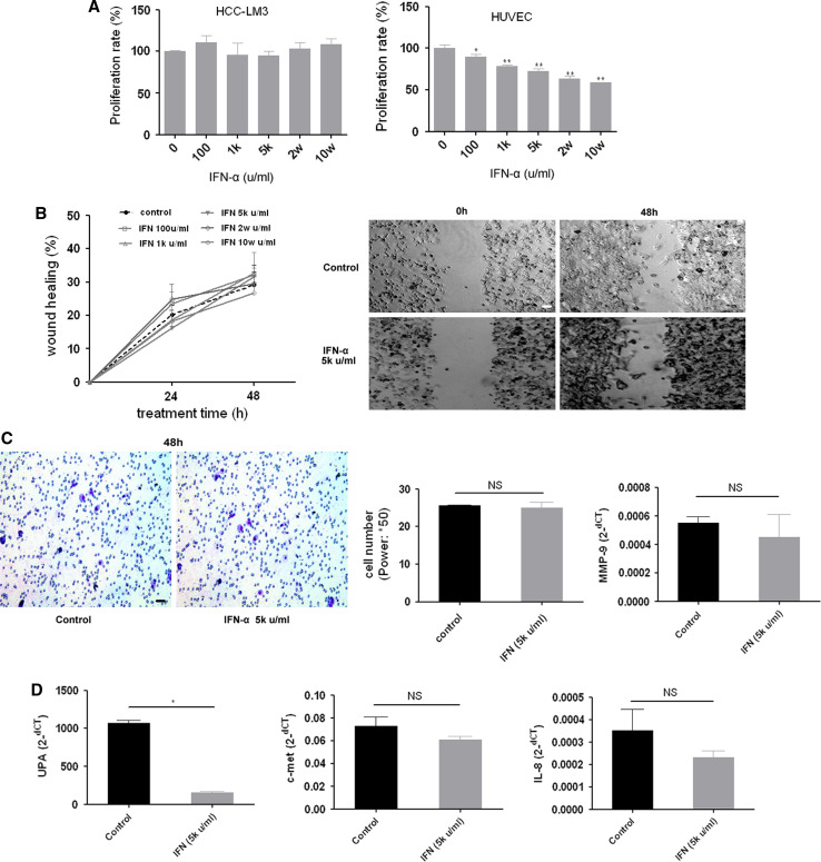
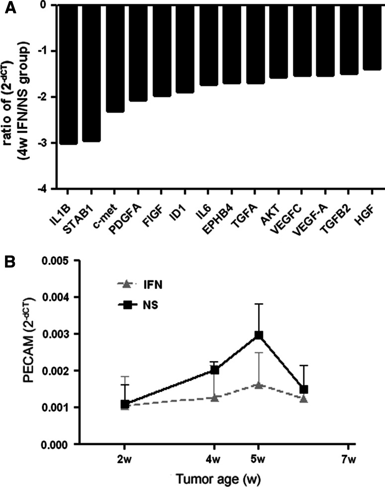
Results: Up to 12 weeks of IFN-α treatment significantly suppressed tumor growth of HCC, but relatively increased the number of circulating tumor cells, which might be due to the enhanced tumor hypoxia as well as up-regulation of metastasis-related genes, such as HIF-1α, c-met, u-PA, PDGF-A, and IL-8. However, IFN-α had no direct effect on migration and invasion of HCC cells.

Conclusions: IFN-α has janus face of consistently suppressing HCC growth, however, promoting tumor metastasis capacity, which is of clinical indication for the scientific administration of IFN-α and the similar antiangiogenesis drugs for their dual effect on tumor growth and metastasis.


Figures

Fig. 1
Fig. 1
Long-term administration of IFN-α inhibited tumor growth and improved survival in mice with orthotopic HCC model. a Tumor growth was significantly inhibited by long-term administration of IFN-α for up to 12 weeks. Tumor size was scrutinized twice a week by B ultrasound. Bars, SEM. b No significant body weight loss was observed between the IFN-α and control groups. c Significantly better overall survival occurred in the long-term administration group than the NS group (log-rank, P = 0.0067). d In the 6-week treatment experiments (orthotopic model using RFP-HCCLM3 cell lines), a much smaller tumor size was observed in the IFN-α group. Pictures of tumor size at 2, 4 and 5 weeks of tumor age. (Upper) NS group; (Lower) IFN-α group
Fig. 2
Fig. 2
Severe tumor hypoxia was induced by IFN-α at a later tumor age. (a and b) Time-course changes of tumor angiogenesis (CD31) and proliferation (Ki-67), measured by immunohistochemistry staining. (c and d) Tumor hypoxia, measured by HIF-1α and pimonidazole staining, in the control group was gradually reduced throughout the tumor growth, whereas IFN-α induced more severe hypoxia than the control group at 7 weeks of tumor age. (e) Immunohistochemistry staining of CD31, ki-67, HIF-1α, and pimonidazole at 7 weeks of tumor age. (Left) The representative immunohistochemistry staining in control (NS) group and IFN-α treatment group. Black bars, 50 μm. (Right) Column represented staining, *** P < 0.001, ** P < 0.01, * P < 0.05
Fig. 3
Fig. 3
IFN-α administration in vitro had no direct effect on tumor proliferation, migration and invasion and the up-regulation of metastasis related factors. a No inhibition of HCC-LM3 cell proliferation by IFN-α was observed in vitro, whereas inhibition on HUVECs by IFN-α was observed in vitro; bars, SEM. ** P < 0.01, * P < 0.05. b IFN-α did not attenuate HCC-LM3 cell migration in vitro for 24 and 48 h. The wound-healing assay was measured by the healing percentage of wound area at the original time point (0 h); bars, SEM. White bars, 50 μm. c Impact on HCC-LM3 motility of IFN-α was determined by Transwell assay. IFN-α did not reduce the HCC-LM3 cell migration for 24 and 48 h, and MMP-9 expression level (real-time PCR) was also not reduced by IFN-α. Black bars, 50um. (D) After incubation of IFN-α for 48 h, IFN-α in vitro did not induce the higher expression of metastasis-related factors such as uPA, c-met, or IL-8 in HCC-LM3 cells, which was confirmed with quantitative real-time PCR. Column was expressed by 2-ΔCT, standardized to human GAPDH. Bars, SEM
Fig. 4
Fig. 4
HCC growth is angiogenesis dependent, and IFN-α could exert its down-regulation of the angiogenesis factors. a IFN-α reduced the angiogenesis peak at the 4 week of tumor age. Comparison of relative RNA levels of angiogenesis—genes between total RNA isolated from tumors harvested from mice treated with IFN-α/control at the 4 week of tumor age. Black bars, relative changes more than 1.5 in ratio to control-treated tumors. Expression levels have been standardized to the probe/primer sets specific for human GAPDH. b Comparison of relative RNA levels of PECAM between total RNA isolated from tumors harvested from both IFN-α-treatment group and control group. Expression levels have been standardized to the probe/primer sets specific for human GAPDH
Fig. 5
Fig. 5
Six-week administration of IFN-α induced up-regulation of metastasis-related genes. a and b Quantitative real-time PCR and Western blotting confirmed the up-regulation of the representative genes compared with the level of homochronous control group. (Left upper) Column was expressed by ratio of 2-ΔCT in the homochronous group. (Left lower) Specific protein expression levels were normalized to the GAPDH protein. *** P < 0.001, ** P < 0.01. (Right) Pictures of Western blotting. c Immunohistochemistry staining confirmed the up-regulation of representative genes in tumor tissue. (Left) The representative staining in the NS group and IFN-α treatment group. Black bars, 50 μm. (Right) Column represented staining, *** P < 0.001, ** P < 0.01, * P < 0.05

Similar articles

Cited by

References

    1. Bix G, Castello R, Burrows M et al (2006) Endorepellin in vivo: targeting the tumor vasculature and retarding cancer growth and metabolism. J Natl Cancer Inst 98:1634–1646 - PubMed
    1. Bristow RG, Hill RP (2008) Hypoxia and metabolism. Hypoxia, DNA repair and genetic instability. Nat Rev Cancer 8:180–192 - PubMed
    1. Chan DA, Giaccia AJ (2007) Hypoxia, gene expression, and metastasis. Cancer Metastasis Rev 26:333–339 - PubMed
    1. Chang YS, Adnane J, Trail PA et al (2007) Sorafenib (BAY 43–9006) inhibits tumor growth and vascularization and induces tumor apoptosis and hypoxia in RCC xenograft models. Cancer Chemother Pharmacol 59:561–574 - PubMed
    1. Fenton BM, Paoni SF (2007) The addition of AG-013736 to fractionated radiation improves tumor response without functionally normalizing the tumor vasculature. Cancer Res 67:9921–9928 - PubMed

Publication types

MeSH terms

Substances

LinkOut - more resources