The role of inflammatory and fibrogenic pathways in heart failure associated with aging
- PMID: 20213186
- PMCID: PMC2920375
- DOI: 10.1007/s10741-010-9161-y
The role of inflammatory and fibrogenic pathways in heart failure associated with aging
Abstract
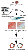
Heart failure is strongly associated with aging. Elderly patients with heart failure often have preserved systolic function exhibiting left ventricular hypertrophy accompanied by a decline in diastolic function. Experimental studies have demonstrated that age-related cardiac fibrosis plays an important role in the pathogenesis of diastolic heart failure in senescent hearts. Reactive oxygen species and angiotensin II are critically involved in fibrotic remodeling of the aging ventricle; their fibrogenic actions may be mediated, at least in part, through transforming growth factor (TGF)-beta. The increased prevalence of heart failure in the elderly is also due to impaired responses of the senescent heart to cardiac injury. Aging is associated with suppressed inflammation, delayed phagocytosis of dead cardiomyocytes, and markedly diminished collagen deposition following myocardial infarction, due to a blunted response of fibroblasts to fibrogenic growth factors. Thus, in addition to a baseline activation of fibrogenic pathways, senescent hearts exhibit an impaired reparative reserve due to decreased responses of mesenchymal cells to stimulatory signals. Impaired scar formation in senescent hearts is associated with accentuated dilative remodeling and worse systolic dysfunction. Understanding the pathogenesis of interstitial fibrosis in the aging heart and dissecting the mechanisms responsible for age-associated healing defects following cardiac injury are critical in order to design new strategies for prevention of adverse remodeling and heart failure in elderly patients.
Figures


References
-
- Chen MA. Heart failure with preserved ejection fraction in older adults. Am J Med. 2009;122:713–723. - PubMed
-
- Thomas S, Rich MW. Epidemiology, pathophysiology, and prognosis of heart failure in the elderly. Clin Geriatr Med. 2007;23:1–10. - PubMed
-
- DeFrances CJ, Cullen KA, Kozak LJ. National hospital discharge survey: 2005 annual summary with detailed diagnosis and procedure data. Vital Health Stat. 2007;13:1–209. - PubMed
-
- Lakatta EG, Levy D. Arterial and cardiac aging: major shareholders in cardiovascular disease enterprises: part II: the aging heart in health: links to heart disease. Circulation. 2003;107:346–354. - PubMed
-
- Kitzman DW, Gardin JM, Gottdiener JS, Arnold A, Boineau R, Aurigemma G, Marino EK, Lyles M, Cushman M, Enright PL. Importance of heart failure with preserved systolic function in patients > or = 65 years of age. CHS research group. Cardiovascular health study. Am J Cardiol. 2001;87:413–419. - PubMed
Publication types
MeSH terms
Substances
Grants and funding
LinkOut - more resources
Full Text Sources
Other Literature Sources
Medical

