Review
. 2010 Jul;460(2):361-74.
doi: 10.1007/s00424-010-0800-x. Epub 2010 Mar 7.

Channelopathies in Cav1.1, Cav1.3, and Cav1.4 voltage-gated L-type Ca2+ channels

Affiliations
Review

Channelopathies in Cav1.1, Cav1.3, and Cav1.4 voltage-gated L-type Ca2+ channels

Jörg Striessnig et al. Pflugers Arch. 2010 Jul.

Abstract

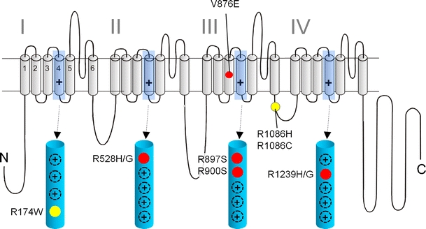
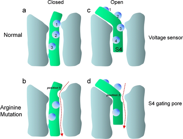
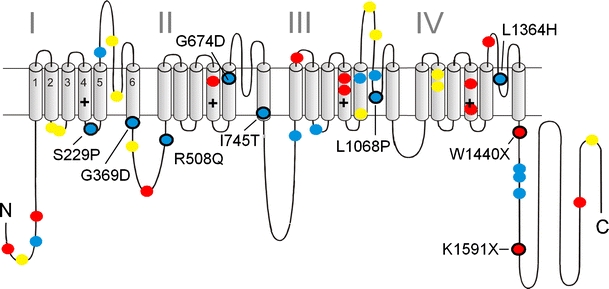
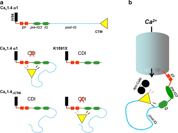
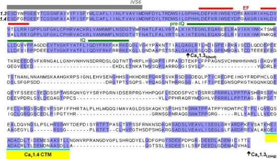
Voltage-gated Ca2+ channels couple membrane depolarization to Ca2+-dependent intracellular signaling events. This is achieved by mediating Ca2+ ion influx or by direct conformational coupling to intracellular Ca2+ release channels. The family of Cav1 channels, also termed L-type Ca2+ channels (LTCCs), is uniquely sensitive to organic Ca2+ channel blockers and expressed in many electrically excitable tissues. In this review, we summarize the role of LTCCs for human diseases caused by genetic Ca2+ channel defects (channelopathies). LTCC dysfunction can result from structural aberrations within their pore-forming alpha1 subunits causing hypokalemic periodic paralysis and malignant hyperthermia sensitivity (Cav1.1 alpha1), incomplete congenital stationary night blindness (CSNB2; Cav1.4 alpha1), and Timothy syndrome (Cav1.2 alpha1; reviewed separately in this issue). Cav1.3 alpha1 mutations have not been reported yet in humans, but channel loss of function would likely affect sinoatrial node function and hearing. Studies in mice revealed that LTCCs indirectly also contribute to neurological symptoms in Ca2+ channelopathies affecting non-LTCCs, such as Cav2.1 alpha1 in tottering mice. Ca2+ channelopathies provide exciting disease-related molecular detail that led to important novel insight not only into disease pathophysiology but also to mechanisms of channel function.


Figures

Fig. 1
Fig. 1
Mutations in Ca2+ channel Cav1.1 α1 subunits identified in patients with HPP-1 and MHS: a folding model of α1-subunits based on hydrophobicity analysis is shown. Plus sign indicates several positive charges in the transmembrane S4 helices within the hydrophobic repeats I–IV. S4 helices and their positively charged residues are shown in the enlarged structures. Together with S1, S2, and S3 helices, they form the four voltage-sensing domains of the channel controlling the opening and closing of a single pore domain formed by S5 and S6 helices together with the connecting linkers. HPP-1 mutations are indicated in red; MHS mutations are shown in yellow. The location of other positive charges in the S4 domains is indicated as black circles (plus sign)
Fig. 2
Fig. 2
Simplified scheme illustrating the membrane potential-dependent conformations of the voltage sensor: only one of the four voltage-sensing domains is illustrated. S4 helices are shown in green, positively charged residues (mostly arginines) as blue spheres. In the closed state, the positively charged S4 helix is pulled inside by the negative resting potential. The outermost arginine residue (1) interacts with residues of other helices forming the voltage-sensing domain (e.g., a key negative charge in S2; [70]) (a). In Shaker K+, Cav1.1, or Nav1.4 channels, a mutation of arginine in position 1 (1) to an uncharged residue (e.g., serine or glycine) opens a new permeation pathway (arrow) as long as the channel is in the closed state (b). Upon depolarization, the S4 helix is driven outward, rotates, and its extracellular portion tilts (c). This movement shifts the arginine in position (3) outward and would close the gating pore induced by a mutation in position 1. The mechanism can account for the depolarizing current observed in muscle cells from HPP-1 patients carrying the Cav1.1 α1 subunit mutations in S4 helices illustrated in Fig. 1 (HPP-1) or analogous mutations in Nav1.4 (HPP-2, not illustrated, [51]). Conversely, whenever the sensor is in the open state, mutation of an arginine in position 3 (3) would enable a gating pore current (d), which would be closed upon repolarization by inward movement of arginine 1. Such a mechanism can explain the depolarization-activated gating pore current conducted by mutant Nav1.4 channels in potassium-sensitive normokalemic periodic paralysis [70]
Fig. 3
Fig. 3
Mutations in Ca2+ channel Cav1.4 α1 subunits identified in patients with CSNB2: a folding model of α1 subunits based on hydrophobicity analysis is shown. Plus sign indicates several positive charges within the transmembrane S4 helices within the hydrophobic repeats I–IV. Position of CSNB2 mutations is indicated. Colors indicate the predicted structural changes: blue, single missense mutations; yellow, in-frame amino acid deletions or insertions; red, truncated protein due to single mutations that introduce stop codons. Black circles refer to mutations that are functionally characterized [–33, 53, 59, 67]
Fig. 4
Fig. 4
Functional CSNB2 mutations in Cav1.4 α1 cause a decreased dynamic range of photoreceptor signaling: the operation range of photoreceptors (between -35 mV (dark) and approximately -55 mV (light) is near the foot of the I Ca activation curve at physiological Ca2+ concentrations to ensure Ca2+ influx necessary for tonic glutamate release (see also text). A hyperpolarizing shift of the current–voltage relationship (IV) is predicted to result in higher glutamate release at a given illumination level, causing a decreased dynamic range of photoreceptor signaling (here shown for mutation K1591X). According to the L-type current IV relationship measured in photoreceptors (black curve [80]), a 13-mV hyperpolarizing shift of the I Ca IV relationship as observed for K1591X [67] would predict a smaller increase of I Ca and exocytosis (predicted: normal ∼50-fold, K1591X ∼3-fold) when moving from the light (-55 mV) to the dark membrane potential (-35 mV)
Fig. 5
Fig. 5
Hypothetical model of Cav1.4 C-terminal modulation. a Motifs previously demonstrated to be important for CaM modulation of other Ca2+ isoforms (red: EF hand; green: pre-IQ regions, IQ domain) are illustrated. In wild-type Cav1.4 channels, the CTM predominantly interacts with a region comprising the EF hand, pre-IQ, and IQ domains and thereby inhibits CDI [67]. The CTM and the post-IQ motif (light blue) are missing in truncation mutant K1591X and therefore intrinsic CDI of Cav1.4 becomes apparent. CDI is present after deletion of the last 122 residues which comprises the CTM. When co-expressed with the truncated channel formula image, the CTM-peptide inhibits CDI and restores wild-type gating properties. This modulation requires the presence of the post-IQ region. In addition, Singh et al. imply a role of the post-IQ motif for voltage-dependent inactivation [67]. b As shown in FRET experiments [70], the Cav1.4 CTM interferes with CaM binding to one or more sites responsible for CaM pre-association (apo-CaM) in intact cells. Therefore, interference with CaM coordination is suggested, the likely mechanism explaining the inhibition of CDI
Fig. 6
Fig. 6
Sequence alignment of C-terminal tails of human Cav1.3 and Cav1.4 L-type channels: a sequence alignment of human Cav1.3 (Genbank accession number EU363339) and Cav1.4 (Genbank accession number AJ224874) α1 subunits is shown. Sequence identity (blue) and gaps (-) are indicated. Regions previously shown to be important for channel modulation by CaM in other voltage-gated Ca2+ channel isoforms are depicted (EF hand, pre-IQ, and IQ domain). The position of long and short Cav1.3 channels is indicated by black arrows (Cav1.3L and Cav1.3S, respectively). Position of the Cav1.4 CTM is given in yellow; - indicates residues absent in this sequence

References

    1. Alseikhan BA, DeMaria CD, Colecraft HM, Yue DT. Engineered calmodulins reveal the unexpected eminence of Ca2+ channel inactivation in controlling heart excitation. Proc Natl Acad Sci U S A. 2002;99:17185–17190. doi: 10.1073/pnas.262372999. - DOI - PMC - PubMed
    1. Badou A, Jha MK, Matza D, Mehal WZ, Freichel M, Flockerzi V, Flavell RA. Critical role for the beta regulatory subunits of Cav channels in T lymphocyte function. Proc Natl Acad Sci U S A. 2006;103:15529–15534. doi: 10.1073/pnas.0607262103. - DOI - PMC - PubMed
    1. Ball SL, Gregg RG. Using mutant mice to study the role of voltage-gated calcium channels in the retina. Adv Exp Med Biol. 2002;514:439–450. - PubMed
    1. Baumann L, Gerstner A, Zong X, Biel M, Wahl-Schott C. Functional characterization of the L-type Ca2+ channel Cav1.4α1 from mouse retina. Invest Ophthalmol Vis Sci. 2004;45:708–713. doi: 10.1167/iovs.03-0937. - DOI - PubMed
    1. Berntson A, Taylor WR, Morgans CW. Molecular identity, synaptic localization, and physiology of calcium channels in retinal bipolar cells. J Neurosci Res. 2003;71:146–151. doi: 10.1002/jnr.10459. - DOI - PubMed

Publication types

MeSH terms

Substances

LinkOut - more resources