. 2010 Jun;40(6):1758-67.
doi: 10.1002/eji.200940158.

Mice lacking C1q or C3 show accelerated rejection of minor H disparate skin grafts and resistance to induction of tolerance

Affiliations
Free PMC article

Mice lacking C1q or C3 show accelerated rejection of minor H disparate skin grafts and resistance to induction of tolerance

Paramita Baruah et al. Eur J Immunol. 2010 Jun.
Free PMC article

Abstract

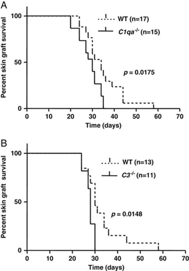
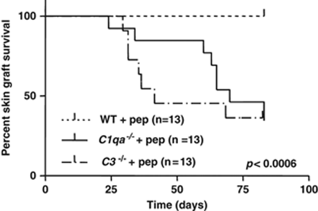
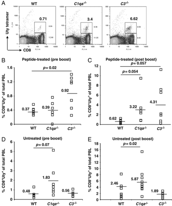
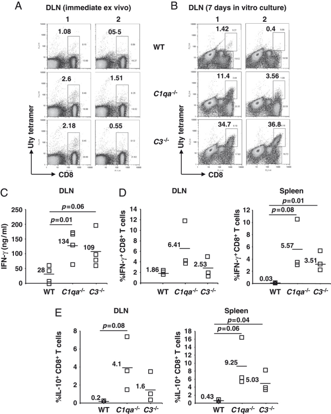
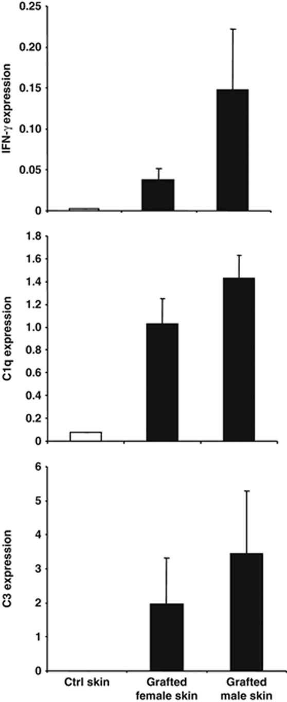
Complement activation is known to have deleterious effects on organ transplantation. On the other hand, the complement system is also known to have an important role in regulating immune responses. The balance between these two opposing effects is critical in the context of transplantation. Here, we report that female mice deficient in C1q (C1qa(-/-)) or C3 (C3(-/-)) reject male syngeneic grafts (HY incompatible) at an accelerated rate compared with WT mice. Intranasal HY peptide administration, which induces tolerance to syngeneic male grafts in WT mice, fails to induce tolerance in C1qa(-/-) or C3(-/-) mice. The rejection of the male grafts correlated with the presence of HY D(b)Uty-specific CD8(+) T cells. Consistent with this, peptide-treated C1qa(-/-) and C3(-/-) female mice rejecting male grafts exhibited more antigen-specific CD8(+)IFN-gamma(+) and CD8(+)IL-10(+) cells compared with WT females. This suggests that accumulation of IFN-gamma- and IL-10-producing T cells may play a key role in mediating the ongoing inflammatory process and graft rejection. Interestingly, within the tolerized male skin grafts of peptide-treated WT mice, IFN-gamma, C1q and C3 mRNA levels were higher compared to control female grafts. These results suggest that C1q and C3 facilitate the induction of intranasal tolerance.


Figures

Figure 1
Figure 1
Accelerated rejection of syngeneic male grafts by C1qa−/− and C3−/− female mice. WT, C1qa−/− and C3−/− female mice were grafted with skin from WT, C1qa−/− and C3−/− male mice, respectively, and graft rejection monitored. (A) Survival of syngeneic male skin grafts transplanted on female WT (n=17) and C1qa−/− (n=15) mice. Data shown are pooled results from three independent experiments (p=0.0175). (B) Survival of syngeneic male grafts transplanted on female WT (n=13) and C3−/− (n=11) mice. Data shown are pooled results from two independent experiments (p=0.0148). Statistical analysis was performed using the log-rank method for survival curves.
Figure 2
Figure 2
Failure of induction of peptide tolerance in C1qa−/− and C3−/− female mice. WT, C1qa−/− and C3−/− female mice were given HYDbUty peptide (100 μg for 3 days) intranasally to induce tolerance. Survival of syngeneic male skin grafts transplanted on peptide-treated female WT (n=13), C1qa−/− (n=13) and C3−/− (n=13) mice (pooled results from two independent experiments) (WT versus C1qa−/− and WT versus C3−/− mice: p<0.0006). Statistical analysis was performed using the log-rank method.
Figure 3
Figure 3
HY antigen-specific Uty+CD8+ T cells in peripheral blood. Female WT, C1qa−/− and C3−/− mice given intranasal peptide 10 days before grafting with syngeneic male skin 10 wk previously were tail-bled before and 7 days after boosting with intraperitoneal male splenocytes. At this time, WT mice were tolerant of their male grafts, while mice in both complement-deficient groups had rejected theirs. PBL were stained with mAb against CD8 and with Uty tetramers. (A) FACS profiles of PBL from one mouse in each group stained ex vivo with anti-CD8 and Uty tetramers 7 days after the intraperitoneal boost. (B, C) Percentage of CD8+Uty+ T cells in PBL of each mouse in the three groups before (B), and after (C), intraperitoneal boosting with male splenocytes. n=6–7 each group. (D, E) Percentage of CD8+Uty+ T cells in PBL of non-peptide-treated WT, C1qa−/− and C3−/− mice subsequently grafted with syngeneic male skin before (n=7 each group) and after intraperitoneal boosting (n=9, 11, 7 respectively; pooled results of two experiments) with male splenocytes. Each symbol represents data from an individual mouse, horizontal bars and the numbers next to the bars indicate the mean (Statistical analysis with Student's t-test).
Figure 4
Figure 4
Ex vivo cytokine responses following intranasal peptide and skin grafting. HYAbDby peptide was administered intranasally to WT, C1qa−/− and C3−/− mice followed by grafting of WT, C1qa−/− and C3−/− male skin respectively ten days later. Following an intraperitoneal boost with male splenocytes 12 wk later, the mice were sacrificed and cells from DLN and spleen harvested. The cells were re-stimulated in vitro with irradiated male splenocytes and stained with anti-CD8 mAb and Uty-tetramer. (A, B) Representative FACS profiles of Uty-tetramer and CD8 on cells of the DLN before and after in vitro restimulation. (C) Cells isolated from DLN were co-cultured with irradiated female or male splenocytes and IFN-γ levels were quantified by ELISA. Each symbol represents data from an individual mouse as the difference in IFN-γ produced following in vitro stimulation with male splenocytes versus female splenocytes. (D) Cells isolated from the DLN and spleen were cultured in vitro with irradiated male splenocytes and restimulated with HYDbUty-peptide-pulsed female splenocytes. IFN-γ-producing CD8+ T cells were quantified by intracellular staining and flow cytometry. (E) Cells from DLN and spleen were cultured in vitro with irradiated male splenocytes and then restimulated with HYDbUty-peptide-pulsed female splenocytes. IL-10-producing CD8+ T cells were quantified by intracellular staining and flow cytometry. (C–E) Results are pooled from two independent experiments; horizontal bars and the numbers next to the bars indicate the mean (statistical analysis with Student's t-test).
Figure 5
Figure 5
Up-regulation of mRNA expression of IFN-γ, C1q and C3 in tolerated male grafts. HYAbDby peptide was administered intranasally to WT female mice followed by grafting of WT male and female skin. The skin grafts were harvested 3 months later and analyzed using quantitative PCR analysis of IFN-γ, C1q and C3 mRNA. Ungrafted female tail skin was used as control. The y-axis represents an arbitrary linear scale of the mean ± SEM mRNA products in skin samples. The results represent pooled data from skin grafts of three mice.

Similar articles

Cited by

References

    1. Kemper C, Atkinson JP. T-cell regulation: with complements from innate immunity. Nat. Rev. Immunol. 2007;7:9–18. - PubMed
    1. Kemper C, Chan AC, Green JM, Brett KA, Murphy KM, Atkinson JP. Activation of human CD4+ cells with CD3 and CD46 induces a T-regulatory cell 1 phenotype. Nature. 2003;421:388–392. - PubMed
    1. Hammerberg C, Katiyar SK, Carroll MC, Cooper KD. Activated complement component 3 (C3) is required for ultraviolet induction of immunosuppression and antigenic tolerance. J. Exp. Med. 1998;187:1133–1138. - PMC - PubMed
    1. Sohn JH, Bora PS, Suk HJ, Molina H, Kaplan HJ, Bora NS. Tolerance is dependent on complement C3 fragment iC3b binding to antigen-presenting cells. Nat. Med. 2003;9:206–212. - PMC - PubMed
    1. Baldwin WM, III, Kasper EK, Zachary AA, Wasowska BA, Rodriguez ER. Beyond C4d: other complement-related diagnostic approaches to antibody-mediated rejection. Am. J. Transplant. 2004;4:311–318. - PubMed

Publication types

MeSH terms