Clinical optical coherence tomography of early articular cartilage degeneration in patients with degenerative meniscal tears
- PMID: 20213801
- PMCID: PMC2972585
- DOI: 10.1002/art.27378
Clinical optical coherence tomography of early articular cartilage degeneration in patients with degenerative meniscal tears
Abstract
Objective: Quantitative and nondestructive methods for clinical diagnosis and staging of articular cartilage degeneration are important to the evaluation of potential disease-modifying treatments in osteoarthritis (OA). Optical coherence tomography (OCT) is a novel imaging technology that can generate microscopic-resolution cross-sectional images of articular cartilage in near real-time. This study tested the hypotheses that OCT can be used clinically to identify early cartilage degeneration and that OCT findings correlate with magnetic resonance imaging (MRI) T2 values and arthroscopy results.
Methods: Patients undergoing arthroscopy for degenerative meniscal tears were recruited under Institutional Review Board-approved protocols. Thirty consecutive subjects completing preoperative 3.0T MRI, arthroscopy, and intraoperative OCT comprised the study group. Qualitative and quantitative OCT results and MRI T2 values were compared with modified Outerbridge cartilage degeneration scores (0-4 scale) assigned at arthroscopy.
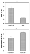
Results: Arthroscopic grades showed cartilage abnormality in 23 of the 30 patients. OCT grades were abnormal in 28 of the 30 patients. Both qualitative and quantitative OCT strongly correlated with the arthroscopy results (P = 0.004 and P = 0.0002, respectively, by Kruskal-Wallis test). Neither the superficial nor the deep cartilage T2 values correlated with the arthroscopy results. The quantitative OCT results correlated with the T2 values in the superficial cartilage (Pearson's r = 0.39, P = 0.03).
Conclusion: These data show that OCT can be used clinically to provide qualitative and quantitative assessments of early articular cartilage degeneration that strongly correlate with arthroscopy results. The correlation between the quantitative OCT values and T2 values for the superficial cartilage further supports the utility of OCT as a clinical research tool, providing quantifiable microscopic resolution data on the articular cartilage structure. New technologies for nondestructive quantitative assessment of human articular cartilage degeneration may facilitate the development of strategies to delay or prevent the onset of OA.
Figures


Similar articles
-
Quantitative Magnetic Resonance Imaging UTE-T2* Mapping of Cartilage and Meniscus Healing After Anatomic Anterior Cruciate Ligament Reconstruction.Am J Sports Med. 2014 Aug;42(8):1847-56. doi: 10.1177/0363546514532227. Epub 2014 May 8. Am J Sports Med. 2014. PMID: 24812196 Free PMC article.
-
Cartilage and meniscus assessment using T1rho and T2 measurements in healthy subjects and patients with osteoarthritis.Osteoarthritis Cartilage. 2010 Nov;18(11):1408-16. doi: 10.1016/j.joca.2010.07.012. Epub 2010 Aug 7. Osteoarthritis Cartilage. 2010. PMID: 20696262 Free PMC article.
-
Optical coherence tomography grading correlates with MRI T2 mapping and extracellular matrix content.J Orthop Res. 2010 Apr;28(4):546-52. doi: 10.1002/jor.20998. J Orthop Res. 2010. PMID: 19834953 Free PMC article.
-
Cartilage and meniscal T2 relaxation time as non-invasive biomarker for knee osteoarthritis and cartilage repair procedures.Osteoarthritis Cartilage. 2013 Oct;21(10):1474-84. doi: 10.1016/j.joca.2013.07.012. Epub 2013 Jul 27. Osteoarthritis Cartilage. 2013. PMID: 23896316 Free PMC article. Review.
-
Articular cartilage lesions in the symptomatic anterior cruciate ligament-deficient knee.Arthroscopy. 2003 Sep;19(7):685-90. doi: 10.1016/s0749-8063(03)00403-1. Arthroscopy. 2003. PMID: 12966374 Review.
Cited by
-
Optical coherence tomography detection of subclinical traumatic cartilage injury.J Orthop Trauma. 2010 Sep;24(9):577-82. doi: 10.1097/BOT.0b013e3181f17a3b. J Orthop Trauma. 2010. PMID: 20736798 Free PMC article.
-
Targeting pro-inflammatory cytokines following joint injury: acute intra-articular inhibition of interleukin-1 following knee injury prevents post-traumatic arthritis.Arthritis Res Ther. 2014 Jun 25;16(3):R134. doi: 10.1186/ar4591. Arthritis Res Ther. 2014. PMID: 24964765 Free PMC article.
-
Correlation of Arthroscopic Grading and Optical Coherence Tomography as Markers of Early Repair and Predictors of Later Healing Evident on MRI and Histomorphometric Assessment of Cartilage Defects Implanted with Chondrocytes Overexpressing IGF-I.Cartilage. 2023 Jun;14(2):210-219. doi: 10.1177/19476035231154508. Epub 2023 Mar 2. Cartilage. 2023. PMID: 36864720 Free PMC article.
-
Changes in viscoelastic properties of articular cartilage in early stage of osteoarthritis, as determined by optical coherence tomography-based strain rate tomography.BMC Musculoskelet Disord. 2019 Sep 6;20(1):417. doi: 10.1186/s12891-019-2789-4. BMC Musculoskelet Disord. 2019. PMID: 31492126 Free PMC article.
-
Novel quantitative imaging for early detection of joint tissue injury to support early treatment strategies.HSS J. 2012 Feb;8(1):54-6. doi: 10.1007/s11420-011-9242-z. Epub 2012 Feb 23. HSS J. 2012. PMID: 23372532 Free PMC article. No abstract available.
References
-
- Radiographic Atlas for Osteoarthritis. Osteoarthritis and Cartilage. 2007;Vol 15 Supplement A
-
- Chu CR, Izzo NJ, Irrgang JJ, Ferretti M, Studer RK. Clinical diagnosis of potentially treatable early articular cartilage degeneration using optical coherence tomography. J Biomed Opt. 2007 Sep–Oct;12(5):051703. - PubMed
-
- Chu CR, Lin D, Geisler JL, Chu CT, Fu FH, Pan Y. Arthroscopic microscopy of articular cartilage using optical coherence tomography. Am J Sports Med. 2004 Apr–May;32(3):699–709. - PubMed
Publication types
MeSH terms
Grants and funding
LinkOut - more resources
Full Text Sources

