. 2010 May;62(5):1523-32.
doi: 10.1002/art.27382.

Selective expression of connective tissue growth factor in fibroblasts in vivo promotes systemic tissue fibrosis

Affiliations

Selective expression of connective tissue growth factor in fibroblasts in vivo promotes systemic tissue fibrosis

Sonali Sonnylal et al. Arthritis Rheum. 2010 May.

Abstract

Objective: Connective tissue growth factor (CTGF) is a cysteine-rich secreted matricellular protein involved in wound healing and tissue repair. Enhanced and prolonged expression of CTGF has been associated with tissue fibrosis in humans. However, questions remain as to whether CTGF expression alone is sufficient to drive fibrosis. This study was undertaken to investigate whether CTGF alone is sufficient to cause fibrosis in intact animals and whether its effects are mediated through activation of transforming growth factor beta (TGFbeta) signaling or through distinct signal transduction pathways.

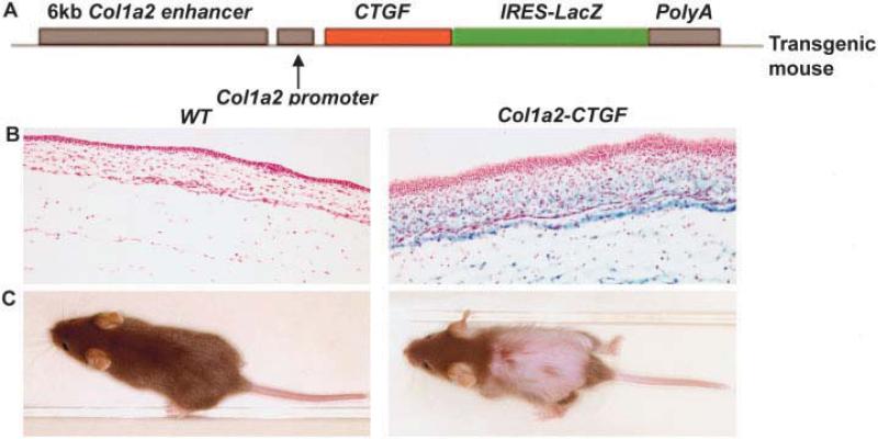
Methods: We generated mice overexpressing CTGF in fibroblasts under the control of the fibroblast-specific collagen alpha2(I) promoter enhancer. Tissues such as skin, lung, and kidney were harvested for histologic analysis. Mouse embryonic fibroblasts were prepared from embryos (14.5 days postcoitum) for biochemical analysis.

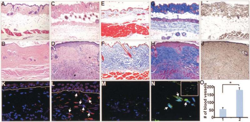
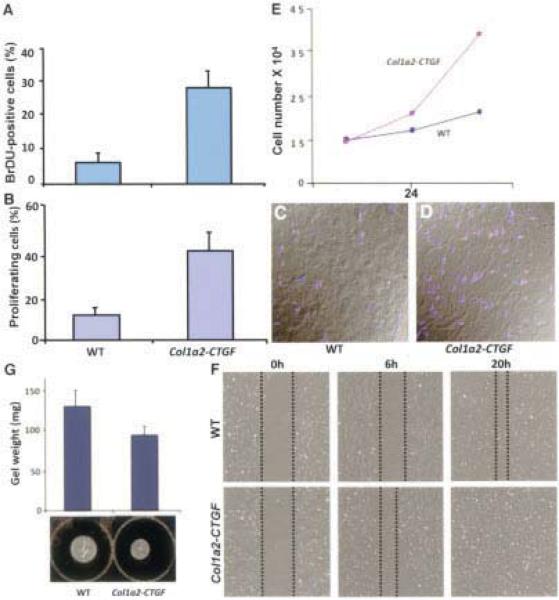
Results: Mice overexpressing CTGF in fibroblasts were susceptible to accelerated tissue fibrosis affecting the skin, lung, kidney, and vasculature, most notably the small arteries. We identified a marked expansion of the myofibroblast cell population in the dermis. RNA analysis of transgenic dermal fibroblasts revealed elevated expression of key matrix genes, consistent with a fibrogenic response. CTGF induced phosphorylation of p38, ERK-1/2, JNK, and Akt, but not Smad3, in transgenic mouse fibroblasts compared with wild-type mouse fibroblasts. Transfection experiments showed significantly increased basal activity of the CTGF and serum response element promoters, and enhanced induction of the CTGF promoter in the presence of TGFbeta.

Conclusion: These results demonstrate that selective expression of CTGF in fibroblasts alone causes tissue fibrosis in vivo through specific signaling pathways, integrating cues from the extracellular matrix into signal transduction pathways to orchestrate pivotal biologic responses relevant to tissue repair and fibrosis.


Figures

Figure 1
Figure 1
Fibroblast-directed overexpression of the connective tissue growth factor (CTGF) gene. A, Schematic representation of the construct in which the coding sequence for the mouse homolog of CTGF, known as Fisp-12, is directed by a fibroblast-specific enhancer and a minimal promoter of the Col1a2 gene. A viral internal ribosome entry site (IRES) sequence–linked lacZ reporter gene and a poly(A) sequence downstream of the CTGF cDNA direct coexpression of this marker from a dicistronic mRNA for identification of transgene expression. B, X-Gal staining of embryos from a Col1a2-CTGF–transgenic mouse and littermate wild-type (WT) control at 15.5 days postcoitum. Intense blue staining of skin was evident only in Col1a2-CTGF–transgenic mouse embryos. C, Gross appearance of 3-week-old WT and Col1a2-CTGF–transgenic mice. The transgenic mouse exhibits severe hair loss from the middle to lower dorsal and upper ventral areas of the body.
Figure 2
Figure 2
Extensive dermal fibrosis in adult Col1a2-CTGF–transgenic mice. A–N, Skin biopsy samples from WT littermate controls (A, C, E, G, I, K, and M) compared with those from adult Col1a2-CTGF–transgenic mice (B, D, F, H, J, L, and N) at 4 weeks of age (A, B, E, and F) and 8 weeks of age (C, D, G–O). Histologic results are representative of skin biopsy samples from 10 mice per group. Compared with WT mice, Col1a2-CTGF–transgenic mice showed pronounced and progressive dermal fibrosis with focal thickening of the epidermis, by hematoxylin and eosin staining (A–D) and Masson's trichrome staining (E–H). Immunochemistry analysis revealed increased accumulation of type I collagen in sections from Col1a2-CTGF–transgenic mice compared with those from WT mice (I and J). Immunofluorescence with CTGF antibody revealed increased CTGF-expressing fibroblasts in Col1a2-CTGF–transgenic mouse skin sections (arrows) compared with WT mouse skin sections (K and L). Increased numbers of myofibroblasts were observed in the papillary and reticular dermis (N and inset in N, respectively) of Col1a2-CTGF–transgenic mouse skin (arrows) compared with WT mouse skin (M). O, Angiogenesis in WT mice (column 1) and Col1a2-CTGF–transgenic mice (column 2), quantified based on numbers of blood vessels. Values are the mean and SD. * = P < 0.001. See Figure 1 for definitions.
Figure 3
Figure 3
Increased fibroblast proliferation in fibrotic skin of Col1a2-CTGF–transgenic mice. A 3-fold increase in bromodeoxyuridine (BrdU)–positive cells was observed in Col1a2-CTGF–transgenic mouse skin sections compared with sections from wild-type (WT) control mice (A). Phosphorylated histone 3 immunostaining of WT (C) and Col1a2-CTGF–transgenic (D) mouse skin sections for evidence of mitotic activity and quantitation of phosphorylated histone 3–positive cells revealed an increase in mitotic activity of cells in the dermis of Col1a2-CTGF–transgenic mice. Quantitative analysis showed that this increase was 2–3-fold (B). Col1a2-CTGF–transgenic mouse embryonic fibroblasts (MEFs) demonstrated 1.3- and 2.6-fold increases in cell number in the first 24 hours and 48 hours, respectively, compared with WT mouse cells (E). Under conditions of serum starvation, scratch assay of MEFs showed increased migration of Col1a2-CTGF–transgenic MEFs by 6 hours (F). Collagen gel contraction assay of WT and Col1a2-CTGF–transgenic MEFs showed increased contraction of the collagen lattice by Col1a2-CTGF–transgenic MEFs (G). Values in A, B, and G are the mean and SD; values in E are the mean.
Figure 4
Figure 4
Pulmonary fibrosis in adult Col1a2-CTGF–transgenic mice. A–D, Lung sections from representative wild-type (WT) mice (A and C) and Col1a2-CTGF–transgenic mice (B and D) exhibited increased staining with Masson's trichrome (A and B) and immunostaining for type I collagen deposition in the alveolar spaces and septa (C and D) in the Col1a2-CTGF–transgenic mice. E, A 4-fold increase in collagen content was observed in Col1a2-CTGF–transgenic mice compared with WT littermates. Values are the mean and SD (n = 5 per group). * = P < 0.05 versus WT mice.
Figure 5
Figure 5
Focal interstitial glomerulosclerosis in Col1a2-CTGF–transgenic mouse kidneys. A–J, Kidney tissue specimens from WT littermate controls (A, C, E, G, and I) compared with those from adult Col1a2-CTGF–transgenic mice (B, D, F, H, and J). Masson's trichrome staining was increased in the basement membrane surrounding the glomeruli of Col1a2-CTGF–transgenic mouse kidneys (A and B). Immunostaining with type IV collagen antibody demonstrated that the accumulated collagen in the interstitium of Col1a2-CTGF–transgenic mice consisted of type IV collagen (C and D). Immunostaining with CTGF antibody showed increased CTGF expression in the glomeruli, surrounding interstitium, in Col1a2-CTGF–transgenic compared with WT mice (E and F). Small blood vessels in the kidney sections stained with Masson's trichrome showed increased collagen deposition around the blood vessels and in the intima in Col1a2-CTGF–transgenic mice compared with WT littermates (G and H). Immunostaining with CD31, a marker for endothelial cells, revealed increased proliferation of endothelial cells in small blood vessels of Col1a2-CTGF–transgenic mice and normal distribution of endothelial cells in WT mice (I and J). K and L, Differential interference contrast overlays of I and J, respectively. See Figure 1 for definitions.
Figure 6
Figure 6
Overexpression of CTGF in fibroblasts causes increased expression of key matrix genes in Col1a2-CTGF–transgenic mouse embryonic fibroblasts (MEFs). A, Northern blotting revealed increased expression levels of genes for CTGF, procollagen α1(I), tissue inhibitors of metalloproteinases 1 and 3, fibronectin, and α-smooth muscle actin in Col1a2-CTGF–transgenic MEFs (lane 2 in A–D) compared with WT MEFs (lane 1 in A–D). B, Western blotting of MEFs showed a corresponding increase in CTGF and type I collagen protein levels in Col1a2-CTGF–transgenic MEFs. C, Western blotting with p-Smad3 antibody revealed no difference in p-Smad protein levels in Col1a2-CTGF–transgenic and WT mouse cells. D, In contrast, constitutive phosphorylation of p38, ERK-1/2, JNK, and Akt was observed in Col1a2-CTGF–transgenic MEFs only. GAPDH, β-actin, and vinculin for total cell extract were used as loading controls; promoter reporter constructs were transfected together with a β-galactosidase vector as an internal control. E, Basal activity levels of serum response element (SRE), CTGF, and CTGF(ΔSmad) promoters were all higher in Col1a2-CTGF–transgenic MEFs than in WT mouse cells. Upon addition of transforming growth factor β1 (TGFβ1), no further activity of the SRE promoter reporter was observed; however, a 3.5-fold increase in CTGF promoter activity occurred. The CTGF promoter with a mutated Smad site showed similar basal activity in the presence and absence of exogenous TGFβ. Values are the mean and SD adjusted level of luciferase (L) or secreted enhanced alkaline phosphatase (SEAP) expression. RLU = relative light units; RSU = relative SEAP units (see Figure 1 for other definitions).

References

    1. Bork P. The modular architecture of a new family of growth regulators related to connective tissue growth factor. FEBS Lett. 1993;327:125–30. - PubMed
    1. Perbal B. NOV (nephroblastoma overexpressed) and the CCN family of genes: structural and functional issues. Mol Pathol. 2001;54:57–79. - PMC - PubMed
    1. Leask A, Abraham DJ. All in the CCN family: essential matricellular signaling modulators emerge from the bunker. J Cell Sci. 2006;119:4803–10. - PubMed
    1. Friedrichsen S, Heuer H, Christ S, Winckler M, Brauer D, Bauer K, et al. CTGF expression during mouse embryonic development. Cell Tissue Res. 2003;312:175–88. - PubMed
    1. Ivkovic S, Yoon BS, Popoff SN, Safadi FF, Libuda DE, Stephenson RC, et al. Connective tissue growth factor coordinates chondrogenesis and angiogenesis during skeletal development. Development. 2003;130:2779–91. - PMC - PubMed

Publication types

MeSH terms