

Structural and biochemical aspects of keratan sulphate in the cornea
- PMID: 20213925
- PMCID: PMC11115788
- DOI: 10.1007/s00018-009-0228-7
Structural and biochemical aspects of keratan sulphate in the cornea
Abstract
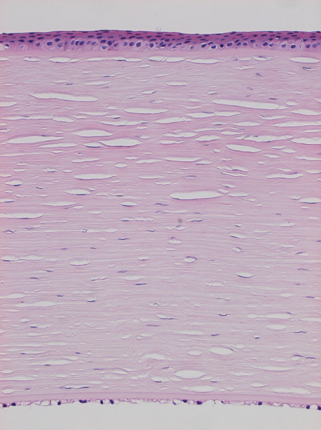
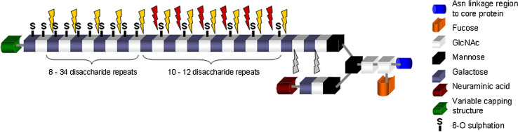
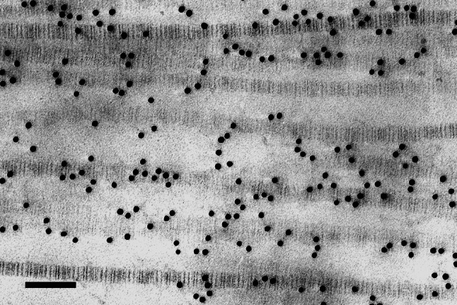
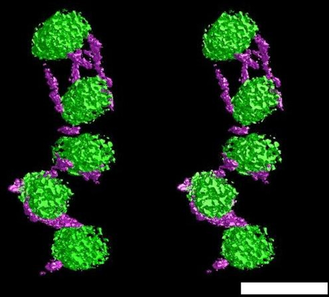
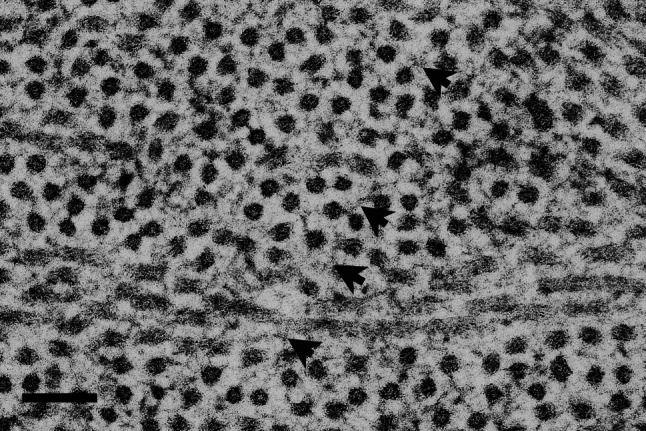
Keratan sulphate (KS) is the predominant glycosaminoglycan (GAG) in the cornea of the eye, where it exists in proteoglycan (PG) form. KS-PGs have long been thought to play a pivotal role in the establishment and maintenance of the array of regularly-spaced and uniformly- thin collagen fibrils which make up the corneal stroma. This characteristic arrangement of fibrils allows light to pass through the cornea. Indeed, perturbations to the synthesis of KS-PG core proteins in genetically altered mice lead to structural matrix alterations and corneal opacification. Similarly, mutations in enzymes responsible for the sulphation of KS-GAG chains are causative for the inherited human disease, macular corneal dystrophy, which is manifested clinically by progressive corneal cloudiness starting in young adulthood.
Figures
Similar articles
-
Role of keratan sulphate (sulphated poly -N-acetyllactosamine repeats) in keratoconic cornea, histochemical, and ultrastructural analysis.Graefes Arch Clin Exp Ophthalmol. 2011 Mar;249(3):413-20. doi: 10.1007/s00417-010-1512-9. Epub 2010 Sep 19. Graefes Arch Clin Exp Ophthalmol. 2011. PMID: 20853116
-
Matrix morphogenesis in cornea is mediated by the modification of keratan sulfate by GlcNAc 6-O-sulfotransferase.Proc Natl Acad Sci U S A. 2006 Sep 5;103(36):13333-8. doi: 10.1073/pnas.0605441103. Epub 2006 Aug 25. Proc Natl Acad Sci U S A. 2006. PMID: 16938851 Free PMC article.
-
A comparison of glycosaminoglycan distributions, keratan sulphate sulphation patterns and collagen fibril architecture from central to peripheral regions of the bovine cornea.Matrix Biol. 2014 Sep;38:59-68. doi: 10.1016/j.matbio.2014.06.004. Epub 2014 Jul 11. Matrix Biol. 2014. PMID: 25019467 Free PMC article.
-
Structure and biological functions of keratan sulfate proteoglycans.EXS. 1994;70:101-22. doi: 10.1007/978-3-0348-7545-5_7. EXS. 1994. PMID: 8298243 Review.
-
Sulfated glycosaminoglycans: their distinct roles in stem cell biology.Glycoconj J. 2017 Dec;34(6):725-735. doi: 10.1007/s10719-016-9732-9. Epub 2016 Oct 6. Glycoconj J. 2017. PMID: 27709407 Review.
Cited by
-
Exploiting Substrate Specificities of 6-O-Sulfotransferases to Enzymatically Synthesize Keratan Sulfate Oligosaccharides.JACS Au. 2023 Oct 13;3(11):3155-3164. doi: 10.1021/jacsau.3c00488. eCollection 2023 Nov 27. JACS Au. 2023. PMID: 38034954 Free PMC article.
-
Molecular engineering of glycosaminoglycan chemistry for biomolecule delivery.Acta Biomater. 2014 Apr;10(4):1705-19. doi: 10.1016/j.actbio.2013.09.039. Epub 2013 Oct 9. Acta Biomater. 2014. PMID: 24121191 Free PMC article. Review.
-
Glycosaminoglycan microarrays for studying glycosaminoglycan-protein systems.Carbohydr Polym. 2024 Jul 1;335:122106. doi: 10.1016/j.carbpol.2024.122106. Epub 2024 Mar 29. Carbohydr Polym. 2024. PMID: 38616080 Free PMC article. Review.
-
Keratan Sulfate Is a Multifunctional Brain Glycosaminoglycan With Instructive Capabilities.J Neurochem. 2025 Aug;169(8):e70208. doi: 10.1111/jnc.70208. J Neurochem. 2025. PMID: 40862528 Free PMC article. Review.
-
Novel mutation in the CHST6 gene causes macular corneal dystrophy in a black South African family.BMC Med Genet. 2016 Jul 20;17(1):47. doi: 10.1186/s12881-016-0308-0. BMC Med Genet. 2016. PMID: 27439461 Free PMC article.
References
-
- Meyer K, Linker A, Davidson EA, Weissmann B. The mucopolysaccharides of bovine cornea. J Biol Chem. 1953;205:611–616. - PubMed
-
- Hassell JR, Cintron C, Kublin C, Newsome DA. Proteoglycan changes during restoration of transparency in corneal scars. Arch Biochem Biophys. 1983;222:362–369. - PubMed
-
- Blochberger TC, Vergnes JP, Hempel J, Hassell JR. cDNA to chick lumican (corneal keratan sulfate proteoglycan) reveals homology to the small interstitial proteoglycan gene family and expression in muscle and intestine. J Biol Chem. 1992;267:347–352. - PubMed
-
- Corpuz LM, Funderburgh JL, Funderburgh ML, Bottomley GS, Prakash S, Conrad GW. Molecular cloning and tissue distribution of keratocan. Bovine corneal keratan sulfate proteoglycan 37A. J Biol Chem. 1996;271:9759–9763. - PubMed
Publication types
MeSH terms
Substances
Grants and funding
- B18021/BB_/Biotechnology and Biological Sciences Research Council/United Kingdom
- BB/D001919/1/BB_/Biotechnology and Biological Sciences Research Council/United Kingdom
- BBS/B/10994/BB_/Biotechnology and Biological Sciences Research Council/United Kingdom
- EY014620/EY/NEI NIH HHS/United States
- R01 EY014620/EY/NEI NIH HHS/United States
LinkOut - more resources
Full Text Sources
