. 2010 Jul;16(7):2133-41.
doi: 10.1089/ten.tea.2009.0670.

Oxygen-controlled three-dimensional cultures to analyze tumor angiogenesis

Affiliations

Oxygen-controlled three-dimensional cultures to analyze tumor angiogenesis

Scott S Verbridge et al. Tissue Eng Part A. 2010 Jul.

Abstract

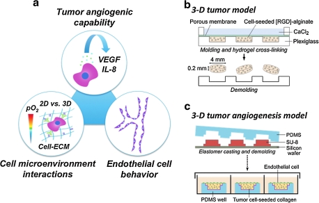
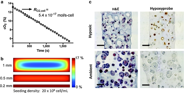
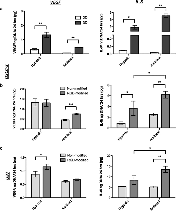
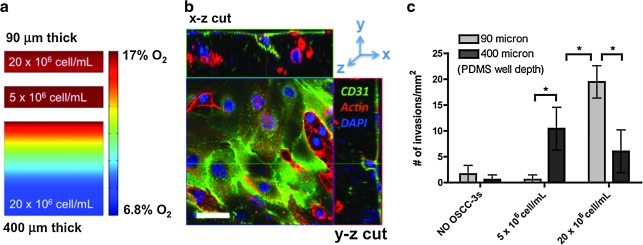
Tumor angiogenesis is controlled by the integrated action of physicochemical and biological cues; however, the individual contributions of these cues are not well understood. We have designed alginate-based microscale tumor models to define the distinct importance of oxygen concentration, culture dimensionality, and cell-extracellular matrix interactions on the angiogenic capability of oral squamous cell carcinoma, and have verified the relevance of our findings with U87 glioblastoma cells. Our results revealed qualitative differences in the microenvironmental regulation of vascular endothelial growth factor (VEGF) and interleukin-8 (IL-8) secretion in three-dimensional (3D) culture. Specifically, IL-8 secretion was highest under ambient conditions, whereas VEGF secretion was highest in hypoxic cultures. Additionally, 3D integrin engagement by RGD-modified alginate matrices increased IL-8 secretion independently of oxygen, whereas VEGF secretion was only moderately affected by cell-extracellular matrix interactions. Using two-dimensional migration assays and a new 3D tumor angiogenesis model, we demonstrated that the resulting angiogenic signaling promotes tumor angiogenesis by increasing endothelial cell migration and invasion. Collectively, tissue-engineered tumor models improve our understanding of tumor angiogenesis, which may ultimately advance anticancer therapies.


Figures

FIG. 1.
FIG. 1.
Study design. (a) To study the pro-angiogenic capability of tumor cells in response to three-dimensional (3D) culture, oxygen (O2) level, and cell–matrix interactions, oral squamous cell carcinoma (OSCC-3) was cultured within unmodified and RGD-modified alginate disks and vascular endothelial growth factor (VEGF) and interleukin-8 (IL-8) secretion was analyzed. Subsequently, the effects of tumor-secreted factors on endothelial cell behavior were determined using a new collagen-based 3D model of invasion. (b) Fabrication of OSCC-3-seeded alginate disks. Alginate solutions were mixed with cells, cast in a micromachined mold, and cross-linked with 60 mM CaCl2. The resulting disks were removed from the mold and used for 3D culture. (c) Three-dimensional tumor angiogenesis models were fabricated by crosslinking of collagen/tumor cell mixtures within poly(dimethylsiloxane) (PDMS) molds that were microfabricated by conventional photolithography techniques. Human umbilical vein endothelial cells were then seeded on the top of these OSCC-3-seeded gels, followed by 3-day culture.
FIG. 2.
FIG. 2.
Characterization of alginate-based 3D tumor models. (a) O2 consumption of OSCC-3 cells seeded within 200-μm-thick alginate disks as quantified by a dissolved O2 meter. (b) Cross-sectional O2 levels of 3D tumor models at day 3 of culture as analyzed using finite element modeling for the measured OSCC-3 consumption rate, and for 200-, 500-, and 1000-μm-thick disks. (c) Histological characterization of alginate disks cultured for 6 days at hypoxic (1% O2) and ambient (18 ± 1% O2) conditions. Hematoxylin and eosin staining indicates uniform cellularity of the developed 3D tumor models. Hypoxyprobe staining identifies hypoxic regions (hypoxic cells appear brown, with blue counterstaining) in cultures maintained at 1% O2, whereas hypoxia was absent in systems cultured at ambient O2. Scale bars represent 50 μm.
FIG. 3.
FIG. 3.
Angiogenic factor secretion. (a) Effect of culture dimensionality (two-dimensional monolayer culture vs. 3D alginate culture) on VEGF and IL-8 secretion by OSCC-3 cells in response to hypoxia and ambient O2 as measured by ELISA of the conditioned medium and normalization to DNA content. (b) Effect of 3D cell–extracellular matrix interactions on VEGF and IL-8 secretion by OSCC-3 cells in response to hypoxia and ambient O2. To prevent and enable 3D integrin engagement, tumor cells were cultured within nonmodified or RGD-modified alginate disks, respectively. The conditioned medium was analyzed as described above. (c) Effect of 3D cell–extracellular matrix interactions on VEGF and IL-8 secretion by U87 cells in response to hypoxia and ambient O2. All 3D medium samples were analyzed after 3 days of culture, whereas two-dimensional medium samples were measured after 2 days to prevent formation of 3D cell aggregates. (*p < 0.05, **p < 0.01, and ***p < 0.001).
FIG. 4.
FIG. 4.
Three-dimensional tumor angiogenesis model. (a) Finite element modeling of O2 distribution in tumor-angiogenesis models of varying cell density and scaffold thickness (90 and 400 μm) to mimic intratumoral spatial variations in O2 levels. Indicated cell numbers (5 × 106 and 20 × 106 cells/mL) represent initial seeding densities, and finite element models account for proliferation by a factor of three after 3 days of culture. (b) Analysis of endothelial cell invasion was performed by confocal imaging subsequent to staining for cell nuclei (blue), actin (red), and the endothelial cell marker CD31 (green). Scale bar represents 50 μm. (c) Number of invading human umbilical vein endothelial cells/mm2 (≥10 μm in depth) quantified for three initial OSCC-3 seeding densities (0, 5 × 106, and 20 × 106 cells/mL) and two collagen scaffold thicknesses (90 and 400 μm) at day 3 of culture (*p < 0.05).

References

    1. Hanahan D. Weinberg R.A. The hallmarks of cancer. Cell. 2000;100:57. - PubMed
    1. Bergers G. Benjamin L.E. Tumorigenesis and the angiogenic switch. Nat Rev Cancer. 2003;3:401. - PubMed
    1. Harris A.L. Hypoxia—a key regulatory factor in tumour growth. Nat Rev Cancer. 2002;2:38. - PubMed
    1. Forsythe J.A. Jiang B.H. Iyer N.V. Agani F. Leung S.W. Koos R.D. Semenza G.L. Activation of vascular endothelial growth factor gene transcription by hypoxia-inducible factor 1. Mol Cell Biol. 1996;16:4604. - PMC - PubMed
    1. Maxwell P.H. Dachs G.U. Gleadle J.M. Nicholls L.G. Harris A.L. Stratford I.J. Hankinson O. Pugh C.W. Ratcliffe P.J. Hypoxia-inducible factor-1 modulates gene expression in solid tumors and influences both angiogenesis and tumor growth. PNAS. 1997;94:8104. - PMC - PubMed

Publication types

MeSH terms

LinkOut - more resources