

Review
doi: 10.1586/era.09.191.
Potential of optical coherence tomography for early diagnosis of oral malignancies
Affiliations
- PMID: 20214513
- PMCID: PMC4038412
- DOI: 10.1586/era.09.191
Item in Clipboard
Review
Potential of optical coherence tomography for early diagnosis of oral malignancies
Expert Rev Anticancer Ther.
2010 Mar.
Abstract
With nearly 1,500,000 new patients diagnosed every year in the USA, cancer poses a considerable challenge to healthcare today. Oral cancer is responsible for a sizeable portion of deaths due to cancer, primarily because it is diagnosed at a late stage when the prognosis is poor. Current methods for diagnosing oral cancer need to be augmented by better early detection, monitoring and screening modalities. A new approach is needed that provides real-time, accurate, noninvasive diagnosis. The results of early clinical trials using in vivo optical coherence tomography for the diagnosis of oral dysplasia and malignancy are encouraging.
Figures
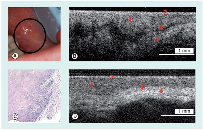
(A) Photograph, (B)
in vivo optical coherence tomography image and (C) hematoxylin and eosin (10×) of dysplastic buccal mucosa. (D)
In vivo optical coherence tomography image of normal buccal mucosa. 1: Stratified squamous epithelium; 2: Keratinized epithelial surface layer; 3: Basement membrane; 4: Submucosa. Reprinted with permission from Wiley-Liss, Inc., a subsidiary of John Wiley & Sons, Inc. [8].
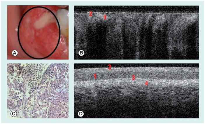
(A) Photograph, (B)
in vivo optical coherence tomography image and (C) hematoxylin and eosin (10×) of buccal mucosa with squamous cell carcinoma. (D)
In vivo optical coherence tomography image of normal buccal mucosa. 1: Stratified squamous epithelium; 2: Keratinized epithelial surface layer; 3: Basement membrane; 4: Submucosa. Reprinted with permission of Wiley-Liss, Inc. a subsidiary of John Wiley & Sons, Inc. [8].
Similar articles
-
Oral Cancer Update: 2017.Northwest Dent. 2017 Jan;96(1):21-3, 25-7. Northwest Dent. 2017. PMID: 30549746 Review. No abstract available.
-
Early detection of premalignant lesions and oral cancer.Otolaryngol Clin North Am. 2011 Feb;44(1):221-9, vii. doi: 10.1016/j.otc.2010.10.002. Otolaryngol Clin North Am. 2011. PMID: 21093631 Review.
-
An update on the clinical pathology of oral precancer and cancer.Dent Update. 2013 Mar;40(2):120-2, 125-6. doi: 10.12968/denu.2013.40.2.120. Dent Update. 2013. PMID: 23600036 Review.
-
[Optical coherence tomography in the evaluation of the oral cavity mucosa. Part II. Benign and malignant diseases].Stomatologiia (Mosk). 2004;83(4):25-32. Stomatologiia (Mosk). 2004. PMID: 15340301 Russian.
-
Exciting new advances in oral cancer diagnosis: avenues to early detection.Head Neck Oncol. 2011 Jul 28;3:33. doi: 10.1186/1758-3284-3-33. Head Neck Oncol. 2011. PMID: 21798030 Free PMC article. Review.
Cited by
-
Tumour Necrosis Factor Alpha (TNF-α) and Oral Squamous Cell Carcinoma.Cancers (Basel). 2023 Mar 19;15(6):1841. doi: 10.3390/cancers15061841. Cancers (Basel). 2023. PMID: 36980727 Free PMC article. Review.
-
Raman spectroscopy in head and neck cancer.Head Neck Oncol. 2010 Oct 5;2:26. doi: 10.1186/1758-3284-2-26. Head Neck Oncol. 2010. PMID: 20923567 Free PMC article. Review.
-
Optical coherence tomography imaging of melanoma skin cancer.Lasers Med Sci. 2019 Mar;34(2):411-420. doi: 10.1007/s10103-018-2696-1. Epub 2018 Dec 11. Lasers Med Sci. 2019. PMID: 30539405 Review.
-
Quantum leap in the diagnosis of oral potentially malignant disorders - A review of literature.Natl J Maxillofac Surg. 2024 Sep-Dec;15(3):360-366. doi: 10.4103/njms.njms_132_23. Epub 2024 Nov 16. Natl J Maxillofac Surg. 2024. PMID: 39830482 Free PMC article. Review.
-
In-vivo Testing of Oral Mucosal Lesions with an In-house Developed Portable Imaging Device and Comparison with Spectroscopy Results.J Fluoresc. 2023 Jul;33(4):1375-1383. doi: 10.1007/s10895-023-03152-z. Epub 2023 Jan 26. J Fluoresc. 2023. PMID: 36701084
References
-
- American Cancer Society . Cancer Facts and Figures. American Cancer Society Report; 2008. pp. 1–4.
-
- Jemal A, Siegel R, Ward E, Murray T, Xu J, Thun J. CA Cancer J. Clin. 2007;57:43–66. - PubMed
-
- Jemal A, Siegel R, Ward E, et al. Cancer statistics. CA Cancer J. Clin. 2008;58(2):71–96. - PubMed
-
- Regezi JA, Sciubba J. Oral Pathology. WB Saunders Co; NY, USA: 1993. pp. 77–90.
-
- Acha A, Ruesga MT, Rodriguez MJ, Pancorbo MA, Aguirre JM. Applications of the oral scraped (exfoliative) cytology in oral cancer and precancer. Oral Surg. Oral Med. Oral Pathol. Oral Radiol. Endodontol - PubMed
Website
-
- US Department of Health and Human Services A national call to action to promote oral health. www.nidr.nih.gov/sgr/nationalcalltoaction.htm