

Phage-displayed combinatorial peptide libraries in fusion to beta-lactamase as reporter for an accelerated clone screening: Potential uses of selected enzyme-linked affinity reagents in downstream applications
- PMID: 20214576
- PMCID: PMC2864580
- DOI: 10.2174/138620710790218258
Phage-displayed combinatorial peptide libraries in fusion to beta-lactamase as reporter for an accelerated clone screening: Potential uses of selected enzyme-linked affinity reagents in downstream applications
Abstract
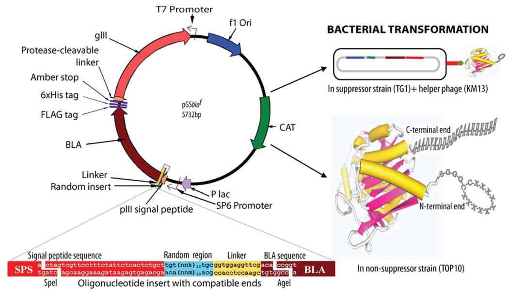
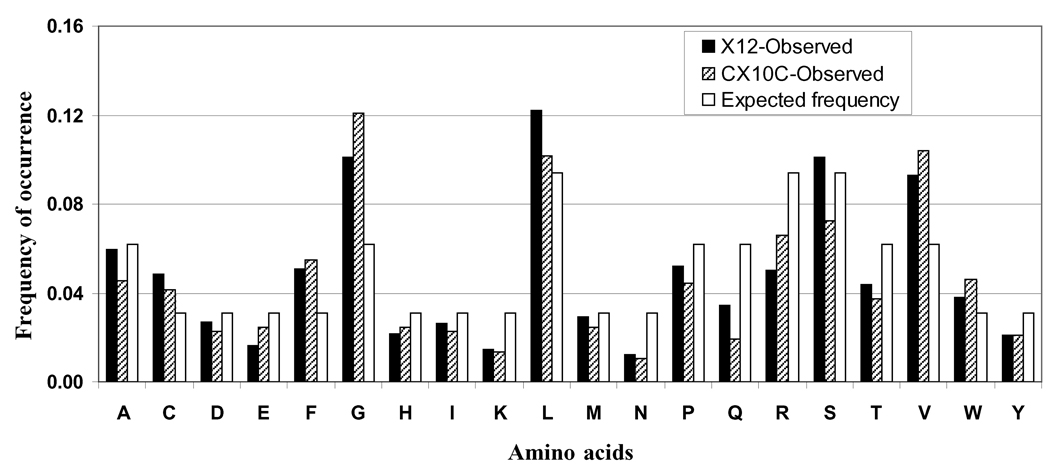
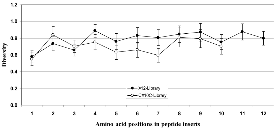
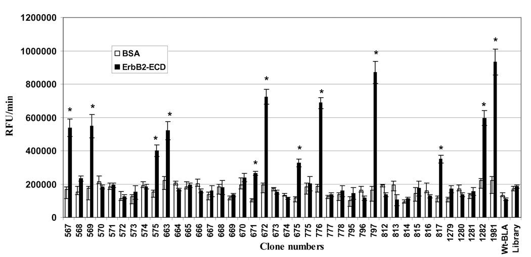
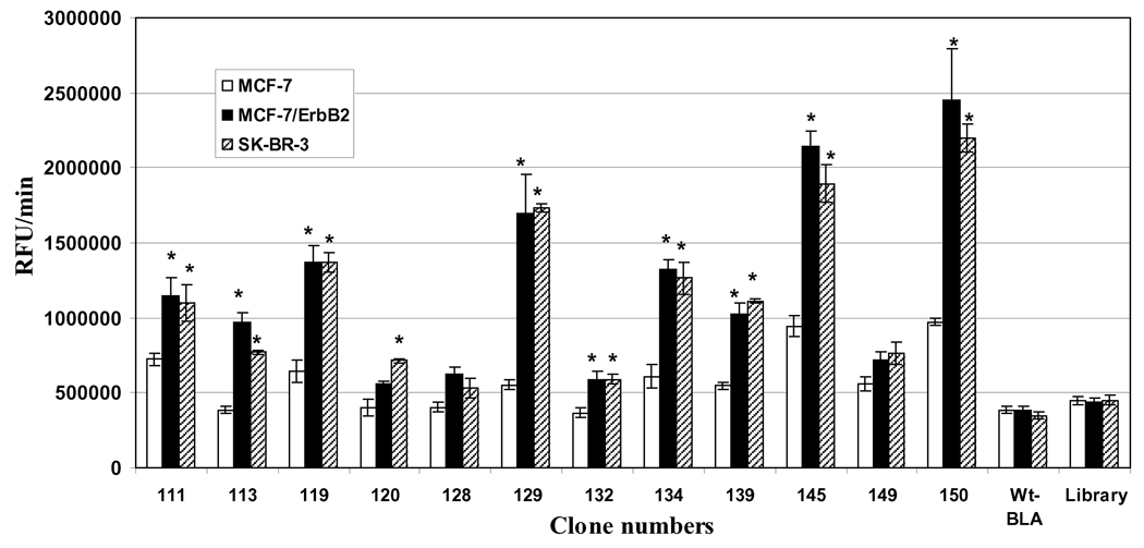
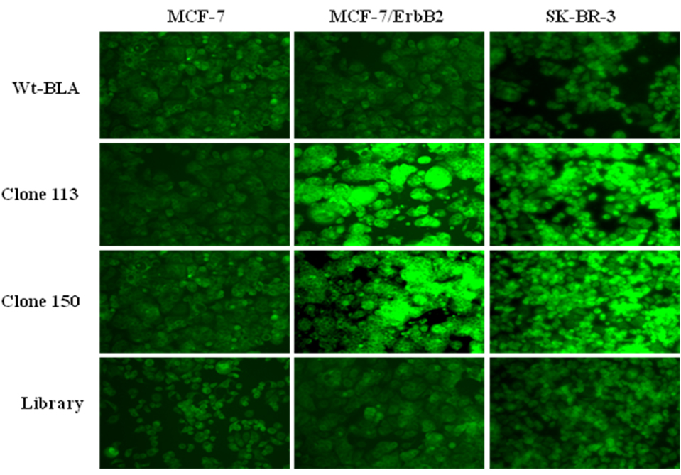
Phage-display selection of combinatorial libraries is a powerful technique for identifying binding ligands against desired targets. Evaluation of target binding capacity of multiple clones recovered from phage display selection to a specific target is laborious, time-consuming, and a rate-limiting step. We constructed phage-display combinatorial peptide libraries in fusion with a beta-lactamase enzyme, which acts as a reporter. Linear dodecapeptide and cysteine-constrained decapeptide libraries were created at the amino-terminus of the Enterobacter cloacae P99 cephalosporinase molecule (P99 beta-lactamase). The overall and positional diversity of amino acids in both libraries was similar to other phage-display systems. The libraries were selected against the extracellular domain of ErbB2 receptor (ErbB2(ECD)). The target-selected clones were already conjugated to an enzyme reporter, therefore, did not require subcloning or any other post-panning modifications. We used beta-lactamase enzyme activity-based assays for sample normalizations and clone binding evaluation. Clones were identified that bound to purified ErbB2(ECD) and ErbB2-overexpressing cell-lines. The peptide sequences of the selected binding clones shared significant motifs with several rationally designed peptide mimetics and phage-display derived peptides that have been reported to bind ErbB2(ECD). beta-Lactamase fusion to peptides saved time and resources otherwise required by the phage-ELISA of a typical phage display screening protocol. The beta-lactamase enzyme assay protocols is a one-step process that does not require secondary proteins, several steps of lengthy incubations, or washings and can be finished in a few minutes instead of hours. The clone screening protocol can be adopted for a high throughput platform. Target-specific beta-lactamase-linked affinity reagents selected by this procedure can be produced in bulk, purified, and used, without any modification, for a variety of downstream applications, including targeted prodrug therapy.
Figures
Similar articles
-
Novel beta-lactamase-random peptide fusion libraries for phage display selection of cancer cell-targeting agents suitable for enzyme prodrug therapy.J Drug Target. 2010 Feb;18(2):115-24. doi: 10.3109/10611860903244181. J Drug Target. 2010. PMID: 19751096 Free PMC article.
-
Developing bifunctional beta-lactamase molecules with built-in target-recognizing module for prodrug therapy: identification of Enterobacter Cloacae P99 cephalosporinase loops suitable for randomization and phage-display selection.J Mol Recognit. 2009 Nov-Dec;22(6):425-36. doi: 10.1002/jmr.957. J Mol Recognit. 2009. PMID: 19437416 Free PMC article.
-
Cancer cell-specific internalizing ligands from phage displayed beta-lactamase-peptide fusion libraries.Protein Eng Des Sel. 2010 Jun;23(6):431-40. doi: 10.1093/protein/gzq013. Epub 2010 Mar 10. Protein Eng Des Sel. 2010. PMID: 20219829 Free PMC article.
-
Phage display biopanning and isolation of target-unrelated peptides: in search of nonspecific binders hidden in a combinatorial library.Amino Acids. 2016 Dec;48(12):2699-2716. doi: 10.1007/s00726-016-2329-6. Epub 2016 Sep 20. Amino Acids. 2016. PMID: 27650972 Review.
-
Biased selection of propagation-related TUPs from phage display peptide libraries.Amino Acids. 2017 Aug;49(8):1293-1308. doi: 10.1007/s00726-017-2452-z. Epub 2017 Jun 29. Amino Acids. 2017. PMID: 28664268 Review.
Cited by
-
Targeting Protein-Protein Interfaces with Peptides: The Contribution of Chemical Combinatorial Peptide Library Approaches.Int J Mol Sci. 2023 Apr 25;24(9):7842. doi: 10.3390/ijms24097842. Int J Mol Sci. 2023. PMID: 37175549 Free PMC article. Review.
-
Bioinformatics resources and tools for phage display.Molecules. 2011 Jan 18;16(1):694-709. doi: 10.3390/molecules16010694. Molecules. 2011. PMID: 21245805 Free PMC article. Review.
References
-
- Bradbury A. Trends Biotechnol. 1999;17:137. - PubMed
-
- de Bruin R, Spelt K, Mol J, Koes R, Quattrocchio F. Nat. Biotechnol. 1999;17:397. - PubMed
-
- Smothers JF, Henikoff S, Carter P. Science. 2002;298:621. - PubMed
-
- Hoogenboom HR, Chames P. Immunol Today. 2000;21:371. - PubMed
-
- Petrenko V. Expert opinion on drug delivery. 2008;5:825. - PubMed
Publication types
MeSH terms
Substances
Grants and funding
LinkOut - more resources
Full Text Sources
Other Literature Sources
Research Materials
Miscellaneous
