

Review
doi: 10.1186/1742-2094-7-18.
Steroid responsive encephalopathy in cerebral amyloid angiopathy: a case report and review of evidence for immunosuppressive treatment
Affiliations
- PMID: 20214781
- PMCID: PMC2846904
- DOI: 10.1186/1742-2094-7-18
Item in Clipboard
Review
Steroid responsive encephalopathy in cerebral amyloid angiopathy: a case report and review of evidence for immunosuppressive treatment
J Neuroinflammation.
.
Abstract
Cerebral amyloid angiopathy (CAA) is a common but often asymptomatic disease, characterized by deposition of amyloid in cerebral blood vessels. We describe the successful treatment of CAA encephalopathy with dexamethasone in a patient with CAA-related inflammation causing subacute progressive encephalopathy and seizures, which is an increasingly recognized subtype of CAA. The two pathological subtypes of CAA-related inflammation are described and a review of the literature is performed concerning immunosuppressive treatment of CAA-related inflammation with special attention to its pathological subtypes. Immunosuppressive therapy appears to be an appropriate treatment for CAA encephalopathy.
Figures
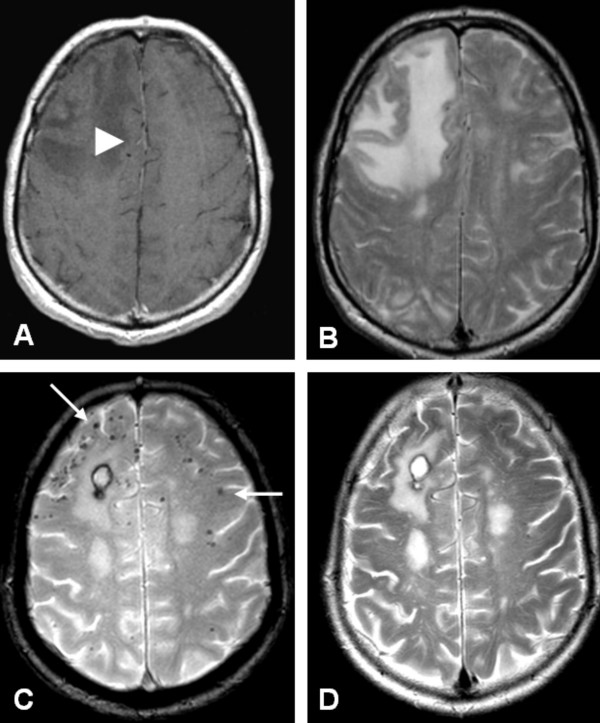
Axial MRI at presentation (A, B) and 3 months after treatment (C, D). A) Contrast enhanced, T1- weighted image shows low signal intensity of the right frontal lobe with minimal enhancement of the white matter (white arrowhead). B) T2-weighted image shows high signal intensity in the right frontal lobe. C) Gradient echo sequence shows subcortical 'black dots', consistent with microbleeds (white arrows), and a small postoperative hematoma after biopsy. D) T2-weighted image after treatment shows a decrease of high signal intensity in the right frontal lobe.
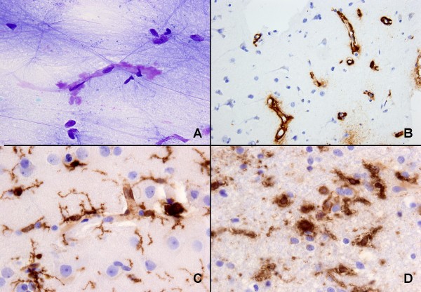
Cytological and histological examination of biopsy. A) Smear slide showing amyloid (metachromatic, purple) around a capillary (toluidin blue stain), B) paraffin-embedded material: extensive amyloid deposition around capillaries in cortex (Aβ immunoreaction), C) reactive gliosis and upregulation of microglia and macrophages in grey matter, D) reactive gliosis, upregulation of microglia and presence of macrophages in white matter (C and D, HLA-DR (CR3/43) immunoreaction).
References
-
- Gilbert JJ, Vinters HV. Cerebral amyloid angiopathy: incidence and complications in the aging brain. I. Cerebral hemorrhage. Stroke. 1983;14:915–23. - PubMed
-
- Greenberg SM, Vonsattel JPG, Stakes JW, Gruber M, Finklestein SP. The clinical spectrum of cerebral amyloid angiopathy: Presentations without lobar hemorrhage. Neurology. 1993;43:2073–2079. - PubMed
Publication types
MeSH terms
Substances
LinkOut - more resources
Full Text Sources
