. 2010 Mar 9:10:28.
doi: 10.1186/1471-213X-10-28.

Cell cycle arrest by a gradient of Dpp signaling during Drosophila eye development

Affiliations

Cell cycle arrest by a gradient of Dpp signaling during Drosophila eye development

Lucy C Firth et al. BMC Dev Biol. .

Abstract

Background: The secreted morphogen Dpp plays important roles in spatial regulation of gene expression and cell cycle progression in the developing Drosophila eye. Dpp signaling is required for timely cell cycle arrest ahead of the morphogenetic furrow as a prelude to differentiation, and is also important for eye disc growth. The dpp gene is expressed at multiple locations in the eye imaginal disc, including the morphogenetic furrow that sweeps across the eye disc as differentiation initiates.

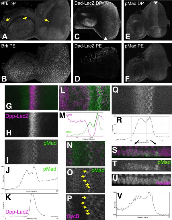
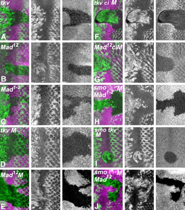
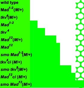
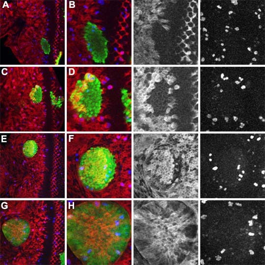
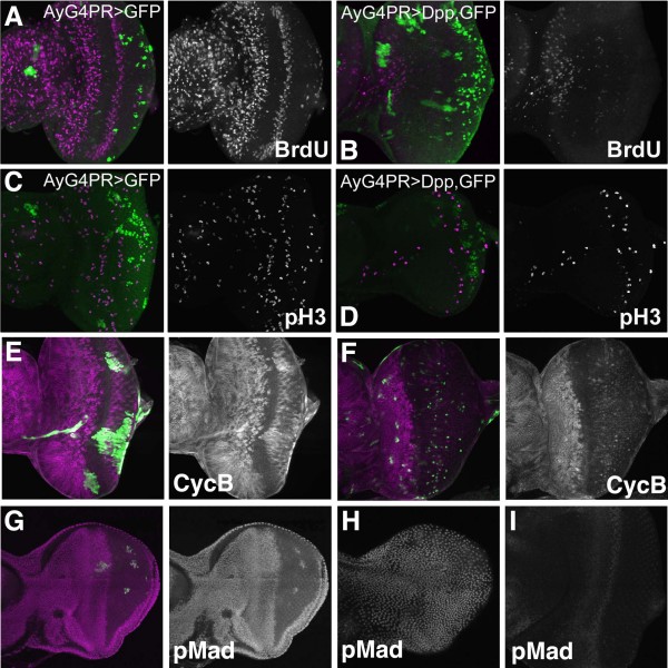
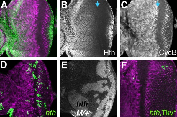
Results: Studies of Brinker and Dad expression, and of Mad phosphorylation, establish that there is a gradient of Dpp signaling in the eye imaginal disc anterior to the morphogenetic furrow, predominantly in the anterior-posterior axis, and also Dpp signaling at the margins of the disc epithelium and in the dorsal peripodial membrane. Almost all signaling activity seems to spread through the plane of the epithelia, although peripodial epithelium cells can also respond to underlying disc cells. There is a graded requirement for Dpp signaling components for G1 arrest in the eye disc, with more stringent requirements further anteriorly where signaling is lower. The signaling level defines the cell cycle response, because elevated signaling through expression of an activated Thickveins receptor molecule arrested cells at more anterior locations. Very anterior regions of the eye disc were not arrested in response to activated receptor, however, and evidence is presented that expression of the Homothorax protein may contribute to this protection. By contrast to activated Thickveins, ectopic expression of processed Dpp leads to very high levels of Mad phosphorylation which appear to have non-physiological consequences.

Conclusions: G1 arrest occurs at a threshold level of Dpp signaling within a morphogen gradient in the anterior eye. G1 arrest is specific for one competent domain in the eye disc, allowing Dpp signaling to promote growth at earlier developmental stages.


Figures

Figure 1
Figure 1
Dpp signaling in the third instar eye-antennal disc. Posterior is to the right and dorsal uppermost in all preparations. (A) Nuclear Brk protein was detected only in parts of the dorsal antennal disc and a few cells at the very anterior of the eye disc proper (arrows); (B) Nuclear Brk protein is strongly detected in peripodial membrane cells. There is a high-to-moderate gradient from ventral-to-dorsal; (C) A positive target of BMP signaling, DadLacZ, was active from 5-6 cell diameters anterior of the morphogenetic furrow and more posteriorly. Arrowhead indicates the morphogenetic furrow. Expression begins slightly earlier (more anteriorly) near the disc margin; (D) DadLacZ was detected in the peripodial epithelium over the ventral antennal disc; (E) Phosphorylated Mad protein is detected in nuclei in a broad domain centered on the MF, extending only slightly more anteriorly at the disc margin, and in a stripe of cells posterior to the furrow. Arrowhead indicates the morphogenetic furrow. Weak labeling of photoreceptor cells in the posterior of the disc is non-specific, since it is unaffected in Mad mutant cells (not shown); (F) Phosphorylated Mad is absent from the peripodial epithelium apart from a few cells dorsally; (G) The Dpp-LacZ transgene is expressed within the morphogenetic furrow (magenta), overlapping the domain of Mad phosphorylation (green). It is possible that Dpp-LacZ might lag behind endogenous Dpp protein; (H) Dpp-LacZ; (I) pMAd; (J) profile plot of the pMad labeling shown in panel I; (K) Profile plot of the Dpp-LacZ shown in panel H. (L) An eye disc labeled with phosphorylated Mad (green) and Cyclin B (magenta) shows that the cells posterior to the furrow where Phospho-Mad levels peak also express the Cyclin B associated with the SMW; (M) Profile plots of the CycB and pMad levels from panel L. (N) A close up of a single confocal z-plane, doubly labeled with CycB and pMad like that shown in panel L. Strongly pMad-positive cells are also labeled with CycB (arrows in panels O, P); (O) pMad labeling from panel N. (P) CycB labeling from panel N. (Q) pMad labeling of an eye disc; (R) Profile plot of the labeling shown in panel Q; (S) A 5 micron strip from panels Q & R, magnified and re-projected to show pMad labeling (green) from the side. Apical disc surface uppermost. Nuclei of all cells are labelled with DRAQ5 (magenta); (T) pMad labeling from panel S; (U) DRAQ5 labeling of all nuclei from panel S; (V) A profile plot from the lateral region of an eye imaginal disc.
Figure 2
Figure 2
G1 arrest requires Dpp and Hh signal reception. All figures show Cyclin B protein (green) in mutant clones and neighboring wild type regions spanning the morphogenetic furrow, with anterior to the left. Homozygous cells are identified by the absence of β-galactosidase (magenta). (A) tkv4; (B)Mad12; (C) Mad1-2; (D) tkv4 in tkv +/+ M background; (E) Mad12 in Mad +/+ M background; (F) tkv4ci94 in tkv ci +/+ + M background; (G) Mad12ci94 in Mad ci +/+ + M background; (H) smo Mad1-2 in smo Mad +/+ + M background; (I) smo3tkv8 in smo tkv +/+ + M background; (J) smo3Mad12 in smo Mad +/+ + M background. Many of these genotypes have also been examined with anti-pH3 labelling to assess mitotic activity, and BrdU incorporation studies to measure S-phase DNA synthesis, confirming the results obtained with CycB labeling in all cases [26](data not shown).
Figure 3
Figure 3
Graded requirement for Dpp and Hh signal transduction. This summary cartoon illustrates in the extent of cell cycle activity near the morphogenetic furrow in various genotypes. Green shading indicate regions where cells have progressed past the G1-S boundary, as indicated by CycB protein levels, for example, whereas white indicates regions where all cells remain in G1. In the case of wild type, shown at the top, all cells remain in G1 from anterior to the morphogenetic furrow until the Second Mitotic Wave starts, after which only the five cells of the photoreceptor preclusters remain in G1. These preclusters appear as the white circles in the Second Mitotic Wave pattern shown at the right of the diagram. Results from 11 other genotypes are summarized in order of severity, with the most severe at the bottom. In each case the approximate extent of the delay in G1 arrest that is observed in mutant clones is indicated. For simplicity, delays in the SMW that result from some genotypes are not included.
Figure 4
Figure 4
Induction of G1 arrest by activated Thickvein. The activated receptor TkvQD was expressed in clones also expressing GFP (green). Cell cycle activity was monitored through labeling for CycB (red) and phospho-H3 (blue). These two markers gave consistent results. 50 TkvQD-expressing pH3-labelled cells in thirteen clones were CycB positive, while 17 pH3-labelled cells in anaphase/telophase were Cyclin B negative, reflecting the metaphase proteolysis of Cyclin B. (A) Near the morphogenetic furrow, close to where wild type cells arrest in G1, TkvQD accelerates arrest of all cells; (B) Higher magnification and separate channels; (C) Clones expressing TkvQD at more anterior locations continued proliferating before arresting about twice as far from the morphogenetic furrow as wild type cells, so that each clone had an anterior, proliferating segment and a posterior, arrested segment; (D) Higher magnification and separated channels; (E) Clone located more anteriorly than that in panel C; (F) higher magnification and separated channels; (G) In the most anterior parts of the eye disc, TkvQDexpression was not sufficient to cause cell cycle arrest; (H) higher magnification and separated channels.
Figure 5
Figure 5
Cell cycle arrest in response to ectopic Dpp. (A) Inducible expression of GFP in clones did not affect cell proliferation in eye discs (BrdU incorporation in magenta); (B) Inducible expression of Dpp in clones almost completely eliminated BrdU incorporation (magenta) throughout the eye disc, antennal disc, and peripodial membrane; (C) Inducible expression of GFP in clones did not affect cell proliferation in eye discs (phospho-H3 labeling in magenta); (D) Inducible expression of Dpp in clones greatly reduced phospho-H3 labeling of mitotic figures; (E) Inducible expression of GFP in clones did not affect CycB expression in eye discs (magenta); (F) Inducible expression of Dpp in clones led to complete loss of CycB expression posterior to the morphogenetic furrow. Anterior to the position where cells normally arrest in G1, where wild type discs contain cells at varied cell cycle stages, ectopic Dpp led to accumulation of CycB in all the arrested cells. CycB in magenta; (G) Even a small proportion of cells expressing Dpp were sufficient for intense Mad phosphorylation (magenta) throughout much of the eye antennal disc. Posterior to the furrow, however, highest levels of pMad were only seen in the cells expressing Dpp themselves; (H) High pMad levels throughout the peripodial membrane of the disc also shown in panel G; (I) pMad labeling in a wild type eye disc processed and recorded in parallel to the disc in panels G and H.
Figure 6
Figure 6
Expression and requirement for homothorax. (A) Homothorax (green) is expressed in the anterior eye disc but repressed anterior to the morphogenetic furrow. Levels gradually reduce, starting while cells are still cycling (CycB in magenta); (B) Hth channel from panel A; Blue arrow shows onset of arrest, as measured by loss of Cyc B (C) CycB channel from panel A; (D) Clones of cells expressing GFP (green) and mutant for hth are recovered in posterior eye regions where cell proliferation stops earlier in development, but rarely recovered in the anterior eye. CycB in magenta; (E) Large hth mutant clones are recovered readily in a heterozygous Minute background. Clones identified by absence of beta-galactosidase labeling; (F) Clones of cells expressing both GFP (green) and TkvQD as well as mutant for hth are recovered very poorly in all locations of the eye disc. CycB in magenta.

Similar articles

Cited by

References

    1. Nellen D, Burke R, Struhl G, Basler K. Direct and long-range action of a DPP morphogen gradient. Cell. 1996;85:357–368. doi: 10.1016/S0092-8674(00)81114-9. - DOI - PubMed
    1. Lecuit T, Brook WJ, Ng M, Callega M, Sun H, Cohen SM. Two distinct mechanisms for long-range patterning by Decapentaplegic in the Drosophila wing. Nature. 1996;381:387–393. doi: 10.1038/381387a0. - DOI - PubMed
    1. Gurdon JB, Mitchell A, Mahony D. Direct and continuous assessment by cells of their position in a morphogen gradient. Nature. 1995;376:520–521. doi: 10.1038/376520a0. - DOI - PubMed
    1. Gurdon JB, Harger P, Mitchell A, Lemaire P. Activin signalling and response to a morphogen gradient. Nature. 1994;371:487–92. doi: 10.1038/371487a0. - DOI - PubMed
    1. Burke R, Basler K. Hedgehog signalling in Drosophila eye and limb development - conserved machinery, divergent roles? Current Opinions in Neurobiology. 1997;7:55–61. doi: 10.1016/S0959-4388(97)80120-1. - DOI - PubMed

Publication types

MeSH terms

Substances

LinkOut - more resources