. 2010 Apr 16;394(4):890-5.
doi: 10.1016/j.bbrc.2010.03.034. Epub 2010 Mar 7.

C-TAK1 interacts with microphthalmia-associated transcription factor, Mitf, but not the related family member Tfe3

Affiliations

C-TAK1 interacts with microphthalmia-associated transcription factor, Mitf, but not the related family member Tfe3

Toni Schwarz et al. Biochem Biophys Res Commun. .

Abstract

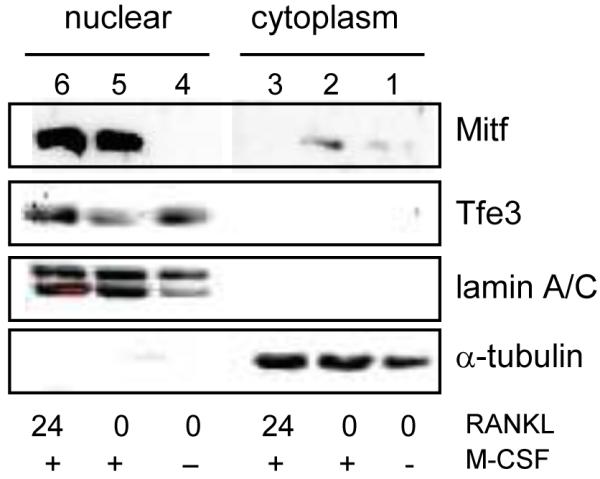
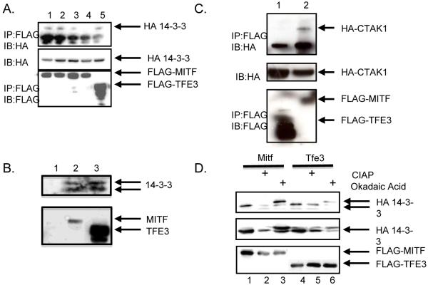
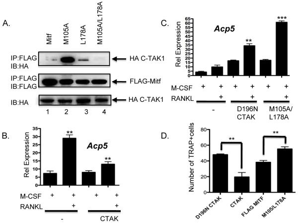
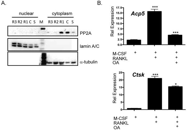
Microphthalmia-associated transcription factor, Mitf, has been shown to be necessary for regulating genes involved in osteoclast differentiation. Previously it was shown by others that Mitf translocates from the cytoplasm to the nucleus upon M-CSF/RANKL signaling in osteoclasts. Mitf's movement is regulated by its interaction with 14-3-3 and the kinase C-TAK1. Here we demonstrate that the related family member, Tfe3, does not shuttle from the cytoplasm to the nucleus and does not interact with C-TAK1. We also demonstrate that overexpression of C-TAK1 inhibits the expression of Acp5 while a kinase dead C-TAK1 or a Mitf mutant that cannot interact with C-TAK1 increased expression of Acp5. Finally, we show that the catalytic subunit of protein phosphatase 2A is up-regulated in osteoclasts with M-CSF/RANKL signaling, indicating a possible mechanism for dephosphorylating Mitf on its 14-3-3 binding site and allowing Mitf to be translocated to the nucleus of osteoclasts.


Figures

Figure 1
Figure 1. Nuclear localization of Mitf and Tfe3
Representative immunoblot of Mitf and Tfe3 lysates from bone marrow derived-osteoclasts. M-CSF and RANKL were removed for 4-6 h, stimulated with M-CSF or M-CSF and RANKL for 24 hours, and immunoblotted.
Figure 2
Figure 2. Mitf and Tfe3 bind to 14-3-3 and C-TAK1
(A) The HA-tagged 14-3-3 construct and the FLAG-tagged Mitf constructs were expressed in 293T cells and complexes from whole-cell lysates were immunoprecipitated with anti-FLAG antibody and analyzed by immunoblot with the indicated antibodies. (B) RAW 264.7 c4 cells were stimulated with 10 ng/mL M-CSF for 24 hours. Cell lysates were immunoprecipitated with an antibody that recognizes IgG (lane 1), Mitf (lane 2) or Tfe3 (lane 3) and analyzed by immunoblot with an antibody that recognizes 14-3-3. (C) HA-tagged C-TAK1 construct and FLAG-tagged Mitf or Tfe3 were expressed in 293T cells and complexes from whole-cell lysates were immunoprecipitated with FLAG antibody and analyzed by immunoblot. (D) FLAG-Mitf or Tfe3 coexpression with HA-14-3-3. Cell lysates were untreated, treated with 20 nM okadaic acid before harvesting, or the whole-cell lysate was treated with 40 U/μl CIAP.
Figure 3
Figure 3. C-TAK1 inhibits Mitf’s ability to activate transcription
(A) The HA-tagged C-TAK1 construct and the FLAG-tagged Mitf point mutants were transfected in 293T cells and complexes from whole-cell lysates were immunoprecipitated with FLAG antibody and analyzed by immunoblot with the indicated antibodies. Real-time RT-PCR from (B) RAW 264.7 c4 cells that were either mock transfected, transiently transfected with full length C-TAK1 **p<0.001 vs. M-CSF mock transfected, **p<0.003 vs. M-CSF C-TAK1 transfected or (C) full length C-TAK1 containing the D196N mutant or Mitf mutant M105A/L178A. Cells were stimulated with M-CSF or M-CSF and RANKL for 5 days. **p<0.0015 vs. RANKL mock transfected, ***p<0.0001 vs. RANKL mock transfected. (D) RAW 264.7c4 cells were either mock-transfected or transfected with wt full-length C-TAK1, D196N C-TAK1, wt FLAG-Mitf or FLAG-Mitf M105/L178A. Cells were cultured in M-CSF and RANKL for 7 d. The number of multinuclear cells (greater than 2 nuclei/cell) was counted and the results of two experiments performed in triplicate are shown. **p<0.007 vs. D196N and **p<0.008 vs. FLAG-Mitf.
Figure 4
Figure 4. Expression of PP2A in osteoclasts
(A) Representative immunoblot of lysates from bone marrow-derived osteoclasts were M-CSF and RANKL was removed for 4-6 h, stimulated with M-CSF or M-CSF and RANKL for up to 72 h and immunoblotted against the catalytic subunit of PP2A, α-tubulin and lamin A/C. (B) Real-time RT-PCR analysis of osteoclasts treated with or without 20 nM oakdaic acid 6 h before harvest for expression of Acp5 ***p<0.0002 vs. M-CSF treated, ***p<0.0005 vs. RANKL treated or cathepsin K, ***p<0.0001 vs. M-CSF treated, *p<0.01 vs. RANKL treated.

Similar articles

Cited by

References

    1. Hemesath TJ, Steingrimsson E, McGill G, Hansen MJ, Vaught J, Hodgkinson CA, Arnheiter H, Copeland NG, Jenkins NA, Fisher DE. microphthalmia, a critical factor in melanocyte development, defines a discrete transcription factor family. Genes Dev. 1994;8:2770–80. - PubMed
    1. Hershey CL, Fisher DE. Mitf and Tfe3: members of a b-HLH-ZIP transcription factor family essential for osteoclast development and function. Bone. 2004;34:689–96. - PubMed
    1. Steingrimsson E, Copeland NG, Jenkins NA. Melanocytes and the microphthalmia transcription factor network. Annu Rev Genet. 2004;38:365–411. - PubMed
    1. Motyckova G, Weilbaecher KN, Horstmann M, Rieman DJ, Fisher DZ, Fisher DE. Linking osteopetrosis and pycnodysostosis: regulation of cathepsin K expression by the microphthalmia transcription factor family. Proc Natl Acad Sci U S A. 2001;98:5798–803. - PMC - PubMed
    1. Luchin A, Purdom G, Murphy K, Clark MY, Angel N, Cassady AI, Hume DA, Ostrowski MC. The microphthalmia transcription factor regulates expression of the tartrate-resistant acid phosphatase gene during terminal differentiation of osteoclasts. J Bone Miner Res. 2000;15:451–60. - PubMed

Publication types

MeSH terms

Substances