

Synaptic characteristics of rostral nucleus of the solitary tract neurons with input from the chorda tympani and glossopharyngeal nerves
- PMID: 20214892
- PMCID: PMC2855769
- DOI: 10.1016/j.brainres.2010.03.003
Synaptic characteristics of rostral nucleus of the solitary tract neurons with input from the chorda tympani and glossopharyngeal nerves
Abstract
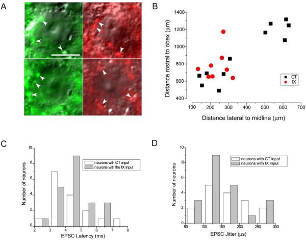
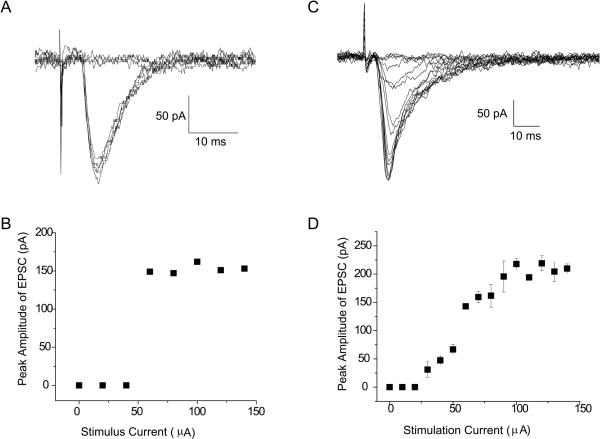
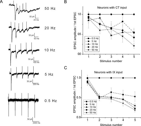
Chorda tympani (CT) and glossopharyngeal (IXth) nerves relay taste information from anterior and posterior tongue to brainstem where they synapse with second order neurons in the rostral nucleus of solitary tract (rNST). rNST neurons monosynaptically connected to afferent gustatory input were identified both by anatomical labeling and synaptic latency measures. Anterograde tracing was used to label the CT and IXth terminal fields, and neurons surrounded by fluorescent neural profiles visualized with differential interference contrast (DIC) optics in horizontal brainstem slices. Anatomically identified neurons were patch-clamped and excitatory postsynaptic currents (EPSCs) evoked by electrically stimulating the solitary tract (ST) under GABA(A) receptor blockade. Monosynaptic connections were confirmed by measures of the standard deviation of synaptic latency (jitter). rNST neurons responded to ST stimulation with either all-or-none or graded amplitude EPSCs. Most (70%) of the rNST neurons with CT input and 30% with IX input responded with all-or-none EPSCs. The remainder of the neurons with CT and IX input responded with increasing EPSC amplitudes to greater intensity stimulus shocks. EPSCs evoked in rNST neurons by increasing shock frequency to both CT and IXth nerves resulted in reduced amplitude EPSCs characteristic of frequency-dependent synaptic depression. Our results suggest that the second order rNST neurons respond to afferent input with different patterns of EPSCs that potentially influence transmission of gustatory information. Frequency-dependent synaptic depression would act as a low pass filter important in the initial processing of gustatory derived sensory messages.
Copyright 2010 Elsevier B.V. All rights reserved.
Figures
Similar articles
-
Characteristics of rostral solitary tract nucleus neurons with identified afferent connections that project to the parabrachial nucleus in rats.J Neurophysiol. 2009 Jul;102(1):546-55. doi: 10.1152/jn.91182.2008. Epub 2009 May 13. J Neurophysiol. 2009. PMID: 19439671 Free PMC article.
-
Synaptic interactions due to convergent input from gustatory afferent fibers in the rostral nucleus of the solitary tract.J Neurophysiol. 1996 Nov;76(5):2919-27. doi: 10.1152/jn.1996.76.5.2919. J Neurophysiol. 1996. PMID: 8930244
-
Chorda tympani nerve stimulation evokes Fos expression in regionally limited neuron populations within the gustatory nucleus of the solitary tract.Brain Res. 2001 Jun 15;904(1):54-66. doi: 10.1016/s0006-8993(01)02449-0. Brain Res. 2001. PMID: 11516411
-
Neural circuits for taste. Excitation, inhibition, and synaptic plasticity in the rostral gustatory zone of the nucleus of the solitary tract.Ann N Y Acad Sci. 1998 Nov 30;855:467-74. doi: 10.1111/j.1749-6632.1998.tb10607.x. Ann N Y Acad Sci. 1998. PMID: 9929640 Review.
-
Neurotransmitter and neuromodulator activity in the gustatory zone of the nucleus tractus solitarius.Chem Senses. 1996 Jun;21(3):377-85. doi: 10.1093/chemse/21.3.377. Chem Senses. 1996. PMID: 8670717 Review.
Cited by
-
Neural coding of taste by simultaneously recorded cells in the nucleus of the solitary tract of the rat.J Neurophysiol. 2012 Dec;108(12):3301-12. doi: 10.1152/jn.00566.2012. Epub 2012 Sep 26. J Neurophysiol. 2012. PMID: 23019002 Free PMC article.
-
The μ-opioid receptor agonist DAMGO presynaptically suppresses solitary tract-evoked input to neurons in the rostral solitary nucleus.J Neurophysiol. 2013 Jun;109(11):2815-26. doi: 10.1152/jn.00711.2012. Epub 2013 Mar 13. J Neurophysiol. 2013. PMID: 23486207 Free PMC article.
-
Inhibitory modulation of optogenetically identified neuron subtypes in the rostral solitary nucleus.J Neurophysiol. 2016 Aug 1;116(2):391-403. doi: 10.1152/jn.00168.2016. Epub 2016 May 4. J Neurophysiol. 2016. PMID: 27146980 Free PMC article.
-
Characteristics of calcium currents in rat geniculate ganglion neurons.J Neurophysiol. 2011 Jan;105(1):224-34. doi: 10.1152/jn.00636.2010. Epub 2010 Nov 10. J Neurophysiol. 2011. PMID: 21068265 Free PMC article.
-
Regulation of Rostral Nucleus of the Solitary Tract Responses to Afferent Input by A-type K+ Current.Neuroscience. 2022 Jul 15;495:115-125. doi: 10.1016/j.neuroscience.2022.05.036. Epub 2022 Jun 2. Neuroscience. 2022. PMID: 35659639 Free PMC article.
References
-
- Andresen MC, Doyle MW, Bailey TW, Jin YH. Differentiation of autonomic reflex control begins with cellular mechanisms at the first synapse within the nucleus tractus solitarius. Braz. J. Med. Biol. Res. 2004;37:549–558. - PubMed
-
- Andresen MC, Yang MY. Dynamics of sensory afferent synaptic transmission in aortic baroreceptor regions of nucleus tractus solitarius. J. Neurophysiol. 1995;74:1518–1528. - PubMed
-
- Araki I, De Groat WC. Unitary excitatory synaptic currents in preganglionic neurons mediated by two distinct groups of interneurons in neonatal rat sacral parasympathetic nucleus. J. Neurophysiol. 1996;76:215–226. - PubMed
-
- Bradley RM. The Role of the Nucleus of the Solitary Tract in Gustatory Processing. CRC Press; Boca Raton: 2006. pp. 1–158. - PubMed
-
- Bradley RM, Sweazey RD. Separation of neuron types in the gustatory zone of the nucleus tractus solitarii based on intrinsic firing properties. J. Neurophysiol. 1992;67:1659–1668. - PubMed
Publication types
MeSH terms
Substances
Grants and funding
LinkOut - more resources
Full Text Sources
