. 2010 Jul;22(7):1063-75.
doi: 10.1016/j.cellsig.2010.02.009. Epub 2010 Mar 7.

Characterization of the expression and regulation of MK5 in the murine ventricular myocardium

Affiliations

Characterization of the expression and regulation of MK5 in the murine ventricular myocardium

Dharmendra Dingar et al. Cell Signal. 2010 Jul.

Abstract

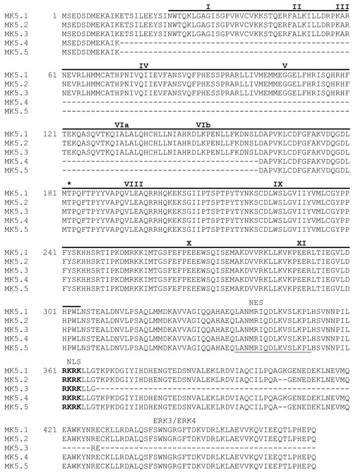
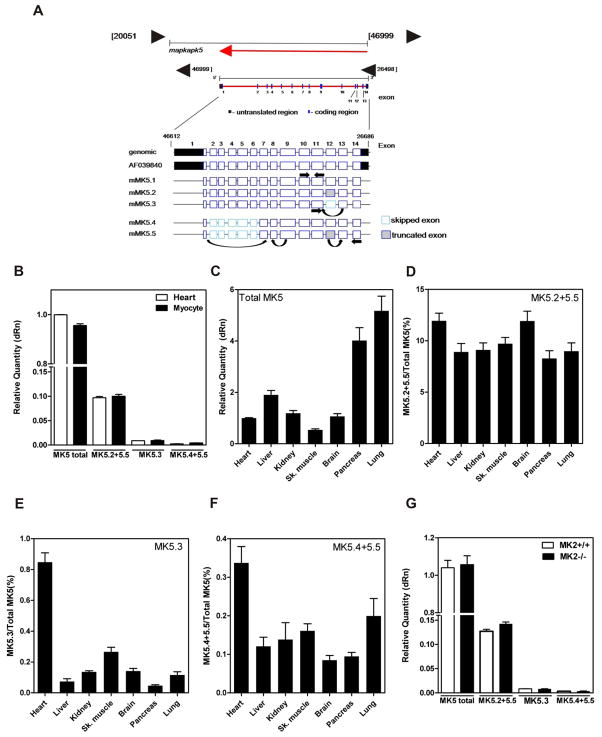
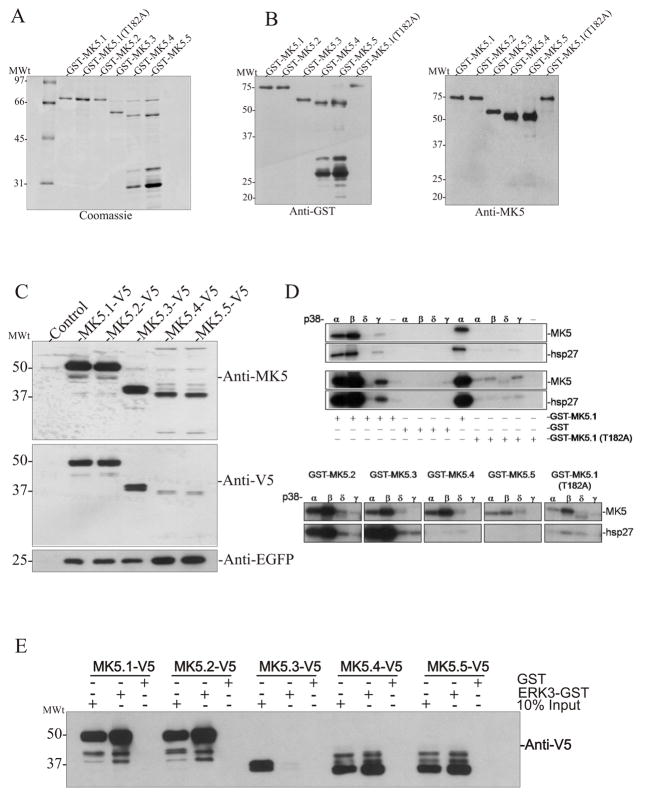
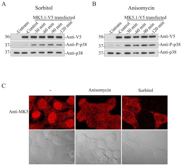
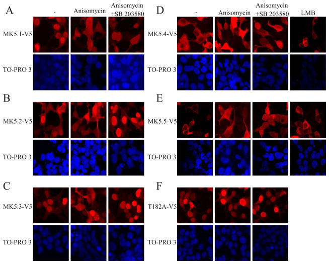
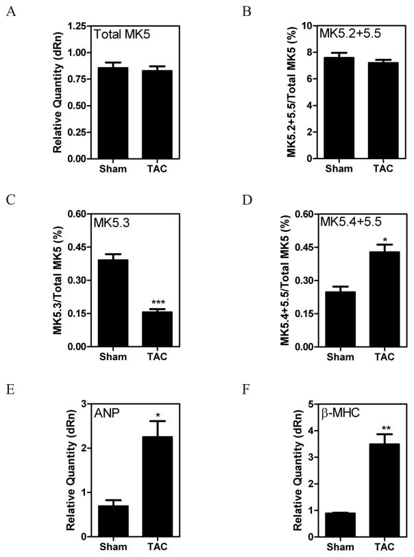
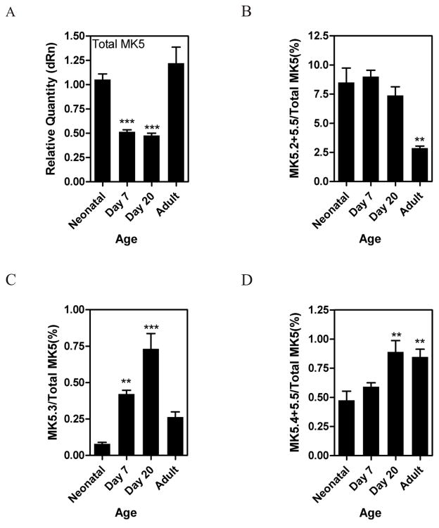
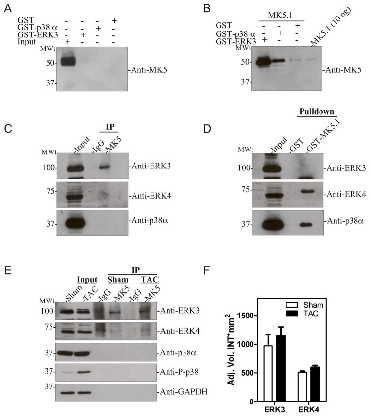
MK5, a member of the MAPK-activated protein kinase family, is highly expressed in the heart. Whereas MK2 and MK3 are activated by p38 MAPK, MK5 has also been shown to be activated by ERK3 and ERK4. We studied the regulation of MK5 in mouse heart. mRNA for 5 splice variants (MK5.1-5.5), including the original form (MK5.1), was detected. MK5 comprises 14 exons: exon 12 splicing was modified in MK5.2, MK5.3, and MK5.5. MK5.2 and MK5.5 lacked 6 bases at the 3'-end of exon 12, whereas MK5.3 lacked exon 12, resulting in a frame shift and premature termination of translation at codon 3 of exon 13. MK5.4 and MK5.5 lacked exons 2-6, encoding kinase subdomains I-VI, and were kinase-dead. All 5 MK5 variants were detected at the mRNA level in all mouse tissues examined; however, their relative abundance was tissue-specific. Furthermore, the relative abundance of variant mRNA was altered both during hypertrophy and postnatal cardiac development, suggesting that the generation or the stability of MK5 variant mRNAs is subject to regulation. When expressed in HEK293 cells, MK5.1, MK5.2 and MK5.3 were nuclear whereas MK5.4 and MK5.5 were cytoplasmic. A p38 MAPK activator, anisomycin, induced the redistribution of each variant. In contrast, MK5 co-immunoprecipitated ERK3, but not ERK4 or p38 alpha, in control and hypertrophying hearts. GST pull-down assays revealed unbound ERK4 and p38 alpha but no free MK5 or ERK3 in heart lysates. Hence, 1) in heart MK5 complexes with ERK3 and 2) MK5 splice variants may mediate distinct effects thus increasing the functional diversity of ERK3-MK5 signaling.


Conflict of interest statement

The authors declare no conflicts of interest.

Figures

Figure 1
Figure 1. Sequence comparison of murine MK5 splice variants
Figure showns the predicted amino acid sequences. Roman numerals above sequence refer to subdomains (I–IX) conserved in all protein kinases. The catalytic domain (residues 22-304) is indicated by a solid line above the sequence. The consensus nuclear export sequence (NES) is underlined with a solid line and the nuclear localization sequence (NES) is in bold letters. Thr-182, the site of phosphorylation by p38 MAPK is indicated by an asterisk. The ERK3/4 binding site is indicated. Dashes represent gaps.
Figure 2
Figure 2. Detection of MK5 variants in murine tissues
(A) Schematic representation of the intron-exon structure of MK5 showing the location of the primers used for quantification of MK5 variant mRNAs. (B) The relative abundance of spliced MK5 mRNAs was measured in total RNA isolated from murine heart and cardiac ventricular myocytes by qPCR. (C) The relative abundance of total MK5 in various murine tissues. Abundance of (D) MK5.2+MK5.5, (E) MK5.3, and (F) MK5.4+MK5.5 mRNA expressed as percentage of total MK5. (G) Detection of MK5 variants in hearts from 12-wk old MK2−/− versus MK2+/+ littermate mice. Shown are the mean ± S.E. (n=3).
Figure 3
Figure 3. Expression of recombinant MK5 splice variants
GST-MK5 fusion proteins were expressed in E. coli and purified by chromatography on glutathione Sepharose. Aliquots of purified protein separated on 10–20% acrylamide-gradient SDS-PAGE and either (A) visualized using Coomassie Brilliant blue R250 stain or (B) transferred to nitrocellulose and probed with anti-GST (left) or anti-MK5 (right) antibodies. (C) HEK293 cells were transfected with the indicated pIRES-MK5-V5-EGFP construct. After 24 h, lysates were prepared, separated on 10–20% acrylamide-gradient SDS-PAGE, transferred to nitrocellulose, and probed with an anti-MK5 (top), anti-V5 (middle), or anti-EGFP (bottom) antibodies. (D) Activated p38 was incubated along with purified GST-MK5, hsp27, and radiolabeled [γ32P]ATP. Proteins were separated on 10–20% SDS-PAGE and 32P incorporation was detected by autoradiography. (E) GST pull-down assays were performed on lysates from HEK293 cells transfected with the indicated expression vectors using ether recombinant ERK3-GST or GST alone. Bound MK5 was detected by immunoblotting using an anti-V5 antibody. Equal expression of the MK5 variants was verified by loading 10% total cell lysate input. Numbers at the left indicate the positions of the molecular mass marker proteins (in kDa).
Figure 4
Figure 4. Activation of p38 MAPK induces translocation of endogenous MK5 in HEK293 cells
HEK293 cells were transfected with pIRES-MK5.1-V5-EGFP. After 24 h cells were serum starved for 6 h and then treated with or without sorbitol (0.3 M) (A) or anisomycin (15 μg/ml) (B) for the indicated times and then lysates were prepared. Activation of p38 was detected by immunoblotting using an anti-phospho p38 antibody. The stability of exogenous MK5 and endogenous p38α during prolonged stress was verified by immunoblotting using anti-V5 and anti-p38α antibodies. (C) Untransfected HEK293 cells were grown for 48 hours, serum starved for 6 h, and then treated with or without sorbitol (0.3 M) or anisomycin (15 μg/ml) for 2 h. Cells were then fixed and MK5 was visualized by staining with an anti-MK5 antibody and an Alexa 555-coupled secondary (anti-mouse) antibody. The upper panels are the fluorescence images whereas the lower panels contain differential interference contrast images of the same cells. Images were acquired using a 63x/1.4 plan-apochromat oil DIC objective lens.
Figure 5
Figure 5. p38 MAPK activation alters the subcellular localization of MK5-V5 variants
HEK293 cells were transfected with (A) pIRES-MK5.1-V5-EGFP, (B) pIRES-MK5.2-V5-EGFP, (C) pIRES-MK5.3-V5-EGFP, (D) pIRES-MK5.4-V5-EGFP, (E) pIRES-MK5.5-V5-EGFP, or (F) pIRES-MK5.1-T182A-V5-EGFP. After 24 h of transfection cells were serum starved for 6 h and then treated with or without anisomycin (15 μg/ml, 2 h), SB203580 (20 μM), or LMB (3 ng/ml, 10 min). Where indicated, cells were preincubated with SB203580 for 10 min prior to the addition of anisomycin. Following treatment, cells fixed and MK5 was visualized by staining with an anti-V5 antibody and Alexa 555-coupled secondary (anti-mouse) antibody (upper panels). Nuclei were visualized by TO-PRO 3 iodide (1.5 μM) staining (lower panels). Several fields of cells were examined and representative images are shown.
Figure 6
Figure 6. Pressure overload-induced hypertrophy alters MK5 splicing
Pressure overload hypertrophy was induced in mice by transverse aortic constriction (TAC). Sham animals underwent the identical surgical procedure, however the aortae were not constricted. After 1 wk of TAC, mice were sacrificed, the hearts isolated, and total RNA isolated. MK5 variants were quantified at the mRNA level by qPCR. GAPDH mRNA was employed as an internal control and the abundance of total MK5 mRNA (A) is expressed relative to GAPDH. The abundance of MK5.2+MK5.5 (B), MK5.3 (C), and MK5.4+MK5.5 (D) mRNAs are expressed as a percentage of total MK5 mRNA. Expression of cardiac fetal genes atrial natriuretic peptide (ANP) (E) and β-myosin heavy chain (β-MHC) (F) are expressed relative to GAPDH. Shown are the mean ± S.E. (n=6). ***, p < 0.001: **, p < 0.01: *, p < 0.05: one-way ANOVA with Newman-Keuls post-hoc analysis.
Figure 7
Figure 7. Regulation of MK5 splicing in heart during postnatal maturation
Mice were sacrificed at birth, 7 d, 20 d, or 8 wk (adult) of age and mRNA levels of MK5 variants quantified by qPCR. The abundance of total MK5 mRNA (A) is expressed relative to GAPDH. The abundance of MK5.2+MK5.5 (B), MK5.3 (C), and MK5.4+MK5.5 (D) mRNAs are expressed as a percentage of total MK5 mRNA. Shown are the mean ± S.E. (n=3). ***, p < 0.001: **, p < 0.01: one-way ANOVA with Newman-Keuls post-hoc analysis.
Figure 8
Figure 8. MK5 interacts with ERK3 in heart
(A) GST pull-down assays were performed on murine ventricular myocardium lysates (2 mg) using either GST-p38α, ERK3-GST, or GST alone. Bound MK5 was detected by immunoblotting using an anti-MK5 monoclonal antibody. As a control, 50 μg lysate (input) was loaded in lane 1. (B) To validate the GST-pull-down assay, lysates (2 mg) were ‘spiked’ with 1 μg of thrombin-cleaved MK5.1 prior to performing the pull-down assay using either GST-p38α, ERK3-GST, or GST alone. Bound MK5 was detected by immunoblotting using an anti-MK5 monoclonal antibody. As a positive control, 10 ng of MK5.1 was loaded onto the gel. (C) Murine ventricular myocardium lysates were prepared and MK5 immunoprecipitated. Purified rabbit IgG was employed in control immunoprecipitations. Co-immunoprecipitated ERK3, ERK4, or p38α was detected by immunoblotting. (D) GST pull-down assays were performed on murine ventricular myocardium lysates (2 mg) using either GST-MK5.1 or GST alone. Bound ERK3, ERK4, or p38α was detected by immunoblotting. As a control, 50 μg of lysate (input) was loaded in lane 1. (E) Pressure-overload hypertrophy was induced in mice by transverse aortic constriction (TAC). Sham animals underwent the identical surgical procedure, however the aortae were not constricted. After 3-d of TAC, mice were sacrificed, the ventricular myocardium isolated, lysates prepared, and MK5 immunoprecipitated. Purified rabbit IgG was employed in control immunoprecipitations. Co-immunoprecipitated ERK3, ERK4, p38α, phospho p38, and GAPDH were detected by immunoblotting. Aliquots (50 μg) of lysate from TAC and sham hearts were included as controls (Input). Results shown are representative of 3 animals in each group. (F) Lysates (50 μg) from 3-d TAC and sham hearts were analyzed by SDS-PAGE and immunoblotting using an anti-ERK3 or anti-ERK4 antibodies. Shown are the mean ± S.E. (n=3).

Similar articles

Cited by

References

    1. Chen Z, Gibson TB, Robinson F, Silvestro L, Pearson G, Xu B, Wright A, Vanderbilt C, Cobb MH. Chem Rev. 2001;101(8):2449–2476. - PubMed
    1. Roux PP, Blenis J. Microbiol Mol Biol Rev. 2004;68(2):320–344. - PMC - PubMed
    1. Kotlyarov A, Neininger A, Schubert C, Eckert R, Birchmeier C, Volk H-D, Gaestel M. Nature Cell Biol. 1999;1:94–97. - PubMed
    1. Gaestel M. Nat Rev Mol Cell Biol. 2006;7(2):120–130. - PubMed
    1. Ludwig S, Engel K, Hoffmeyer A, Sithanandam G, Neufeld B, Palm D, Gaestel M, Rapp UR. Mol Cell Biol. 1996;16(12):6687–6697. - PMC - PubMed

Publication types

MeSH terms

Substances