. 2010 Apr 30;285(18):13326-36.
doi: 10.1074/jbc.M109.077297. Epub 2010 Mar 9.

Controlled expression of branch-forming mannosyltransferase is critical for mycobacterial lipoarabinomannan biosynthesis

Affiliations

Controlled expression of branch-forming mannosyltransferase is critical for mycobacterial lipoarabinomannan biosynthesis

Chubert B C Sena et al. J Biol Chem. .

Abstract

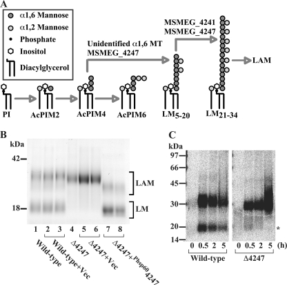
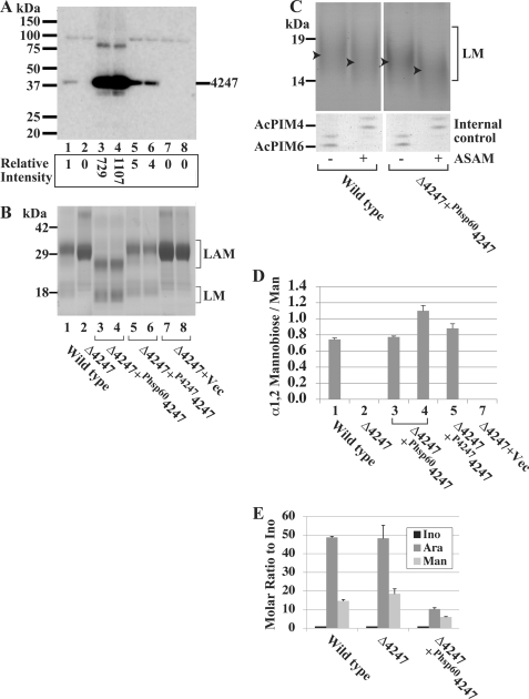
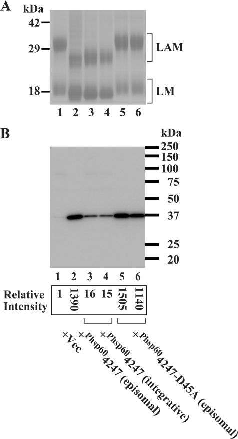
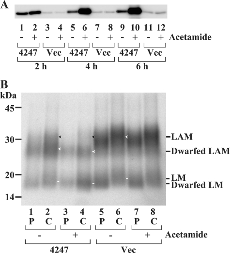
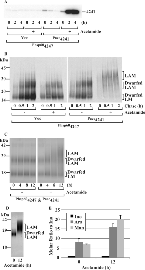
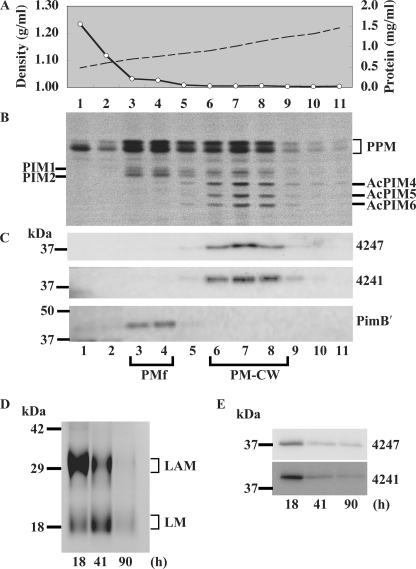
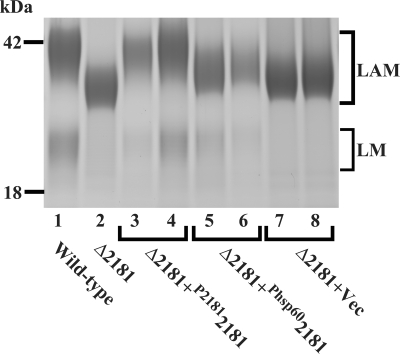
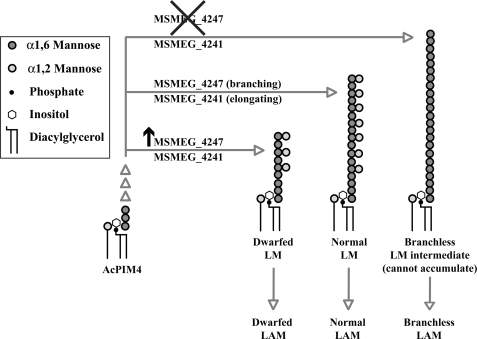
Lipomannan (LM) and lipoarabinomannan (LAM) are phosphatidylinositol-anchored glycans present in the mycobacterial cell wall. In Mycobacterium smegmatis, the mannan core of LM/LAM constitutes a linear chain of 20-25 alpha1,6-mannoses elaborated by 8-9 alpha1,2-monomannose side branches. At least two alpha1,6-mannosyltransferases mediate the linear mannose chain elongation, and one branching alpha1,2-mannosyltransferase (encoded by MSMEG_4247) transfers monomannose branches. An MSMEG_4247 deletion mutant accumulates branchless LAM and interestingly fails to accumulate LM, suggesting an unexpected role of mannose branching for LM synthesis or maintenance. To understand the roles of MSMEG_4247-mediated branching more clearly, we analyzed the MSMEG_4247 deletion mutant in detail. Our study showed that the deletion mutant restored the synthesis of wild-type LM and LAM upon the expression of MSMEG_4247 at wild-type levels. In striking contrast, overexpression of MSMEG_4247 resulted in the accumulation of dwarfed LM/LAM, although monomannose branching was restored. The dwarfed LAM carried a mannan chain less than half the length of wild-type LAM and was elaborated by an arabinan that was about 4 times smaller. Induced overexpression of an elongating alpha1,6-mannosyltransferase competed with the overexpressed branching enzyme, alleviating the dwarfing effect of the branching enzyme. In wild-type cells, LM and LAM decreased in quantity in the stationary phase, and the expression levels of branching and elongating mannosyltransferases were reduced in concert, presumably to avoid producing abnormal LM/LAM. These data suggest that the coordinated expressions of branching and elongating mannosyltransferases are critical for mannan backbone elongation.


Figures

FIGURE 1.
FIGURE 1.
The phenotype of MSMEG_4247 deletion mutant (ΔMSMEG_4247) cannot be restored by introduction of the episomal MSMEG_4247 overexpression vector. A, proposed pathway of PIM, LM, and LAM biosynthesis. Note that AcPIM4 is a proposed branch point from which the LM/LAM biosynthesis pathways diverge. The mannosyltransferase (MT) that mediates the initial elongation of the α1,6-mannose chain to produce LM5–20 has not been identified. The positions of the α1,2 side branches are hypothetical. Only the products and key intermediates are shown, and tetra-acylated PIMs are not shown for simplicity. B, LM/LAM from ΔMSMEG_4247 and its complemented strains were analyzed by SDS-PAGE and visualized by carbohydrate staining. Wild-type (lanes 1–3) and ΔMSMEG_4247 mutant (lanes 4–8) were transfected with an empty vector (lanes 2, 3, 5, and 6, pYAB251) or an episomal Phsp60MSMEG_4247 vector (lanes 7 and 8, pYAB250). Loading was adjusted for equal cell pellet equivalents. C, [3H]mannose metabolic labeling of LM and LAM in the ΔMSMEG_4247 mutant strain. Cells were pulsed with [3H]mannose for 15 min and chased with excess non-radioactive mannose for up to 5 h. LM and LAM profiles were analyzed by SDS-PAGE and fluorography. Transient synthesis of LM is indicated by an asterisk.
FIGURE 2.
FIGURE 2.
Controlled expression of MSMEG_4247 is critical for the restoration of LM/LAM biosynthesis. A, MSMEG_4247 expression examined by Western blotting using anti-MSMEG_4247 antibody. Lane 1, wild type; lane 2, ΔMSMEG_4247; lanes 3 and 4, two clones of ΔMSMEG_4247+Phsp60MSMEG_4247 (pYAB250); lanes 5 and 6, two clones of ΔMSMEG_4247+P4247MSMEG_4247 (pYAB247); lanes 7 and 8, two clones of ΔMSMEG_4247 transfected with empty integrative vector (pYAB184). Loading was adjusted to 5 μg of protein/lane except for lanes 3 and 4, in which 1 μg of protein was loaded per lane. Relative intensities were calculated taking different protein loadings into account. B, LM/LAM profiles of MSMEG_4247 deletion mutants complemented by various expression vectors. LM/LAM were analyzed by SDS-PAGE and visualized by carbohydrate staining. Lanes are arranged in the same order as in A. Faint doublet bands slightly above the 18 kDa marker seen in all lanes are protein contaminants. C, LM from wild-type or ΔMSMEG_4247+Phsp60MSMEG_4247 was treated with or without α1,2-mannosidase (ASAM), analyzed by SDS-PAGE using Tris-Tricine gel (15–20%) to improve the separation, and visualized by carbohydrate staining. Peak of LM in each lane was determined from the intensity profile and is indicated by an arrowhead. PIMs were included as internal controls and showed specific and complete digestion of the terminal two α1,2-mannoses of AcPIM6, producing AcPIM4. D, LM/LAM extracts were acetolyzed, and released mannose and α1,2-mannobiose were detected by HPAEC. Molar ratios of α1,2-mannobiose to mannose, measured in triplicate, are shown as averages with S.D. values. Column numbers specify the samples analyzed in the corresponding lanes in A and B. E, LAM was purified by electroelution and hydrolyzed by 2 m trifluoroacetic acid. Released carbohydrates were quantified by HPAEC. Data are presented as molar ratio relative to inositol. Averages of triplicate measurements with S.D. values are shown.
FIGURE 3.
FIGURE 3.
Overexpression of MSMEG_4247 reduces the sizes of LM and LAM in wild-type cells. A, LM/LAM profiles of wild-type cells transfected with various Phsp60-driven expression vectors were analyzed by SDS-PAGE and visualized by carbohydrate staining. Lane 1, empty vector (pHBJ334); lane 2, episomal vector to express MSMEG_4247 (pYAB143); lanes 3 and 4, integrative vector to express MSMEG_4247 (pYAB243); lanes 5 and 6, episomal vector to express MSMEG_4247 D45A mutant (pYAB254). B, MSMEG_4247 expression examined by Western blotting using anti-MSMEG_4247 antibody. Lanes are arranged in the same order as in A. Loading was adjusted to 5 μg of protein/lane for lanes 1, 3, and 4 or 0.25 μg/lane for lanes 2, 5, and 6. Relative intensities were calculated, taking different protein loadings into account.
FIGURE 4.
FIGURE 4.
Acetamide-induced overexpression of MSMEG_4247 prevents the maturation of LM/LAM. A, acetamide induction of MSMEG_4247 examined by Western blotting using anti-MSMEG_4247 antibody. Wild-type cells transfected with either the acetamide-inducible MSMEG_4247 expression vector (4247) (transfected with pYAB246) or empty vector (Vec) (transfected with pYAB040) were incubated with (+) or without (−) acetamide for the indicated period to induce MSMEG_4247 expression. B, metabolic labeling with [3H]mannose. Cells were incubated with (+) or without (−) acetamide for 4 h prior to radiolabeling. Cells were pulsed (P) for 15 min and then chased (C) in the presence of excess non-radioactive mannose for 25 min. The white arrowheads and lines indicate the peaks of LAM and LM, respectively, in lanes 2, 4, and 6, as determined by the intensity profiles shown in supplemental Fig. S4. The black arrowheads indicate the position of normal LAM as determined by the white arrowhead in lane 6.
FIGURE 5.
FIGURE 5.
Dwarfing effect of MSMEG_4247 overexpression on LM/LAM can be competed by overexpression of MSMEG_4241. A, acetamide induction of MSMEG_4241 examined by Western blotting using anti-MSMEG_4241 antibody. The MSMEG_4247 overexpression mutant was transfected with either acetamide-inducible MSMEG_4241 expression vector (Pace4241) (transfected with pYAB262) or empty vector (Vec) (transfected with pJAM2) and incubated with (+) and without (−) acetamide for the indicated period to induce MSMEG_4241 expression. The loading was adjusted to 5 μg of protein/lane. B, metabolic labeling with [3H]mannose. Cells were preincubated with (+) or without (−) acetamide for 4 h, pulsed for 15 min with [3H]mannose, and then chased in the presence of excess non-radioactive mannose for up to 2 h. C, changes in total LM/LAM profiles after induction of MSMEG_4241 expression in MSMEG_4247 overexpression mutant. LM/LAM were separated by SDS-PAGE and visualized by carbohydrate staining. D, dwarfed LAM at 0 h and LAM-like species at 12 h after acetamide induction, shown in C, were purified by electroelution, and the purities were confirmed by SDS-PAGE and carbohydrate staining. E, compositional analysis of LAM-like species produced after MSMEG_4241 overexpression. Trifluoroacetic acid-hydrolyzed carbohydrates were quantified by HPAEC. Data are presented as molar ratio relative to inositol. Averages of triplicate measurements with S.D. values are shown.
FIGURE 6.
FIGURE 6.
Subcellular localization and growth phase-dependent expression of MSMEG_4247 and MSMEG_4241 in wild-type cells. A, sucrose density (dashed line) and protein concentration (open circle) profiles of wild-type cell lysate fractionated by a density sedimentation. B, PIM biosynthetic activities of each fraction measured by GDP-[3H]mannose radiolabeling. PPM, polyprenol-phosphate-mannose. C, Western blotting using anti-MSMEG_4247 (top), anti-MSMEG_4241 (middle), or anti-PimB′ (bottom) antibodies. D, growth phase-dependent changes in LM/LAM levels of wild type cells grown in Middlebrook 7H9 broth. Culture was initiated by 1:100 dilution of a confluent starter culture, and aliquots were collected at the indicated time points, which correspond to logarithmic (18 h), early stationary (41 h), and late stationary (90 h) phases. Purified LM/LAM were separated by SDS-PAGE and visualized by carbohydrate staining. Loading was adjusted for equal cell pellet equivalents. E, growth phase-dependent changes in levels of MSMEG_4247 and MSMEG_4241 detected by Western blotting using anti-MSMEG_4247 or anti MSMEG_4241 antibodies. Loading was adjusted to 15 μg of protein/lane.
FIGURE 7.
FIGURE 7.
LM/LAM profiles of M. tuberculosis Rv2181 deletion mutants transfected with various expression vectors. LM/LAM were extracted, analyzed by SDS-PAGE, and visualized by carbohydrate staining. Lane 1, wild type; lane 2, ΔRv2181 mutant; lanes 3 and 4, ΔRv2181 mutant transfected with pYAB228, an integrative expression vector carrying Rv2181, including 242 bp of upstream sequence; lanes 5 and 6, ΔRv2181 mutant transfected with pYAB230, an integrative expression vector carrying Rv2181 driven by Phsp60; lanes 7 and 8, ΔRv2181 mutant transfected with pYAB184, an integrative empty vector control.
FIGURE 8.
FIGURE 8.
A model of LM/LAM biosynthesis involving elongating (MSMEG_4241) and branching (MSMEG_4247) mannosyltransferases. The lack of MSMEG_4247 results in the lack of LM and accumulation of branchless LAM. In contrast, overexpression of MSMEG_4247 results in dwarfed LM and dwarfed LAM. The positions of α1,2-mannose branches are hypothetical. Although our data are consistent with LM being a precursor of LAM biosynthesis, the precursor-product relationship of LM and LAM remains to be proved. The structure of LM intermediate in ΔMSMEG_4247 mutant is hypothetical, based on the structure of LAM accumulating in the mutant.

Similar articles

Cited by

References

    1. Brennan P. J. (2003) Tuberculosis 83, 91–97 - PubMed
    1. Briken V., Porcelli S. A., Besra G. S., Kremer L. (2004) Mol. Microbiol. 53, 391–403 - PubMed
    1. Alderwick L. J., Birch H. L., Mishra A. K., Eggeling L., Besra G. S. (2007) Biochem. Soc. Trans. 35, 1325–1328 - PubMed
    1. Khoo K. H., Dell A., Morris H. R., Brennan P. J., Chatterjee D. (1995) Glycobiology 5, 117–127 - PubMed
    1. Lee Y. C., Ballou C. E. (1964) J. Biol. Chem. 239, 1316–1327 - PubMed

Publication types

MeSH terms

LinkOut - more resources