. 2010 Apr 30;285(18):13372-9.
doi: 10.1074/jbc.M109.083089. Epub 2010 Mar 9.

Pyk2 activation triggers epidermal growth factor receptor signaling and cell motility after wounding sheets of epithelial cells

Affiliations

Pyk2 activation triggers epidermal growth factor receptor signaling and cell motility after wounding sheets of epithelial cells

Ethan R Block et al. J Biol Chem. .

Abstract

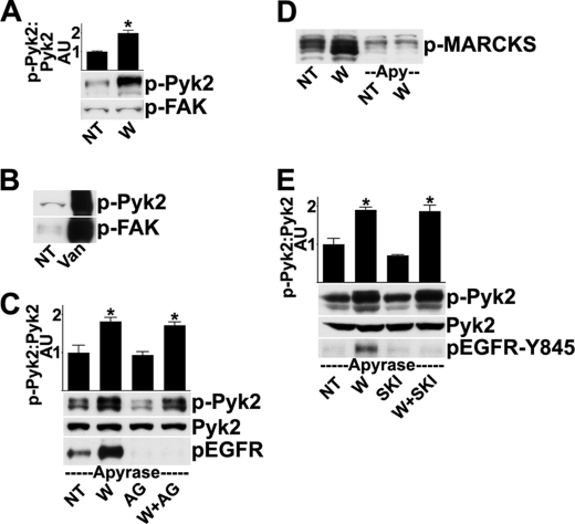
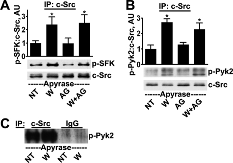
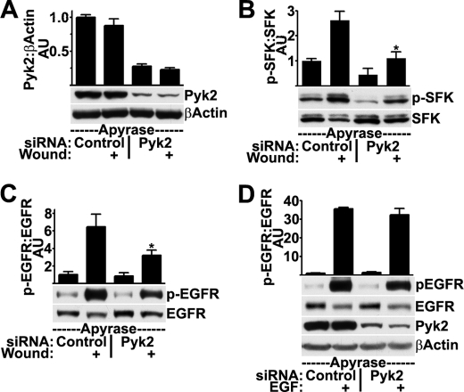
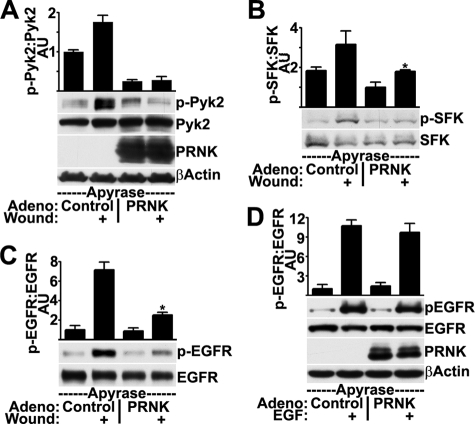
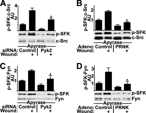
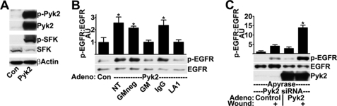
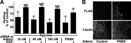
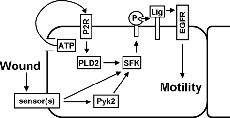
Activation of the epidermal growth factor receptor (EGFR) is a key signaling event that promotes cells to move and cover wounds in many epithelia. We have previously shown that wounding activates the EGFR through activation of the Src family kinases (SFKs), which induce proteolytic shedding of epidermal growth factor-like ligands from the cell surface. A major goal in wound healing research is to identify early signals that promote motility, and here we examined the hypothesis that members of the focal adhesion kinase family are upstream activators of the SFKs after wounding. We found that focal adhesion kinase is not activated by wounding but that a different family member, Pyk2 (PTK2B/RAFTK/CAKbeta), is activated rapidly and potently. Pyk2 interaction with c-Src is increased after wounding, as determined by co-immunoprecipitation experiments. Disruption of Pyk2 signaling either by small interfering RNA or by expression of a dominant negative mutant led to inhibition of wound-induced activation of the SFKs and the EGFR, and conversely, overexpression of wild-type Pyk2 stimulated SFK and EGFR kinase activities in cells. In wound healing studies, Pyk2 small interfering RNA or dominant negative inhibited cell migration. These results show that activation of Pyk2 is an early signal that promotes wound healing by stimulating the SFK/EGFR signaling pathway.


Figures

FIGURE 1.
FIGURE 1.
Pyk2, not FAK, is activated rapidly after wounding. A, HCLE cells were given no treatment (NT) or were wounded (W) and incubated for 5 min, and cell extracts were analyzed by Western blotting for Pyk2 phosphorylated on Tyr-402 (p-Pyk2) or for FAK phosphorylated on Tyr-397 (p-FAK). Due to various splicing and post-translational events, the antibody for phosphorylated Pyk2 detects multiple bands, all of which are used in analysis. The ratio of phospho-Pyk2/Pyk2 (not shown) was determined by densitometry. *, significant differences from unwounded groups (p < 0.001). Columns are means, and error bars are S.D. B, as a positive control for the antibodies, 100 nm Na3VO4 was added to the cells for 30 min prior to analysis (Van). C, in the presence of 25 units/ml apyrase, cells received no additional treatment (NT) or were wounded (W) and incubated for 5 min. Where indicated, cells were treated with 100 nm tyrphostin AG 1478 (AG) for 30 min prior to and during wounding. Western blots were probed for phospho-Pyk2 and were then stripped and reprobed for total levels of Pyk2 and EGFR phosphorylated on Tyr-1173 (p-EGFR). The ratio of phospho-Pyk2/Pyk2 was determined by densitometry. *, significant differences from unwounded groups (p < 0.001). D, cells were treated as indicated, and Western blots were probed for phosphorylated myristoylated alanine-rich C-kinase substrate (p-MARCKS), which is a marker of protein kinase C activity (29, 30). E, cells were treated as in C, except 1 μm of Src kinase inhibitor-I (SKI) was added for 30 min prior to and during wounding, where indicated. The blots were analyzed with an antibody that recognizes the EGFR phosphorylated on Tyr-845 (p-EGFR-Y845). Densitometry was performed as in C. Apy, apyrase.
FIGURE 2.
FIGURE 2.
c-Src is activated and interacts with Pyk2 after wounding. Cells received no treatment (NT) or were wounded (W) and incubated for 5 min, all conditions in the presence of 25 units/ml apyrase. Where indicated, cells were treated with 100 nm tyrphostin AG 1478 (AG) for 30 min prior to and during wounding. The extracts were precipitated with c-Src antibodies, which removed ∼90% of the c-Src present in the extracts (not shown), and the precipitates were subjected to Western blotting for SFK phosphorylated on Tyr-419 (p-SFK) and subsequently for total levels of c-Src (A) and Pyk2 phosphorylated on Tyr-402 (p-Pyk2) and total levels of c-Src (B). Ratios were determined by densitometry. *, significant differences from unwounded groups (p < 0.01). Columns are means, and error bars are S.D. C, lysates were subjected to immunoprecipitation (IP) with anti-c-Src antibodies or with non-immune mouse IgG; immunoprecipitates were subjected to Western blotting for phospho-Pyk2. Overexposed film shows the absence of signal from the non-immune control.
FIGURE 3.
FIGURE 3.
Knockdown of Pyk2 with siRNA inhibits SFK and EGFR activation after wounding but not after stimulation with EGF. HCLE cells transfected with 10 nm control or Pyk2 siRNA were treated with apyrase and were wounded as indicated. Western blots were probed for total levels of Pyk2 and β-actin (A), SFK phosphorylated (p-SFK) and non-phosphorylated (SFK) on Tyr-419 (B), and EGFR phosphorylated on Tyr-1173 (p-EGFR) and total levels of EGFR (C). Ratios were determined by densitometry. *, significant decrease from wounded controls (p < 0.001). D, HCLE cells transfected with control or Pyk2 siRNA were treated with 10 ng/ml EGF for 5 min where indicated prior to Western blotting and densitometry. Columns are means, and error bars are S.D.
FIGURE 4.
FIGURE 4.
Expression of a Pyk2 dominant negative mutant inhibits SFK and EGFR activation after wounding but not after stimulation with EGF. HCLE cells infected with control adenovirus or adenovirus coding for PRNK were incubated with apyrase and were wounded as indicated. Western blots were probed for Pyk2 phosphorylated on Tyr-402 (p-Pyk2) and total levels of Pyk2, PRNK, and β-actin (A), SFK phosphorylated (p-SFK) and non-phosphorylated (SFK) on Tyr-419 (B), and EGFR phosphorylated on Tyr-1173 (p-EGFR) and total levels of EGFR (C). Ratios were determined by densitometry. *, significant decrease from wounded controls (p < 0.001). D, HCLE cells infected with control adenovirus or adenovirus coding for PRNK were treated with 10 ng/ml EGF for 5 min where indicated prior to Western blotting and densitometry. Columns are means, and error bars are S.D.
FIGURE 5.
FIGURE 5.
Disruption of Pyk2 signaling by siRNA or PRNK inhibits wound-induced activation of c-Src and Fyn. HCLE cells transfected with 10 nm control or Pyk2 siRNA (A and C) or infected with control adenovirus or adenovirus coding for PRNK (B and D) were treated with apyrase and were wounded as indicated. c-Src immunoprecipitates were immunoblotted for SFK phosphorylated on Tyr-419 (p-SFK) and for total levels of c-Src (A and B). Fyn immunoprecipitates were immunoblotted for SFK phosphorylated on Tyr-419 and for total levels of Fyn (C and D). Ratios were determined by densitometry. *, significant decrease from wounded controls (p < 0.001). Columns are means, and error bars are S.D.
FIGURE 6.
FIGURE 6.
Overexpression of wild-type Pyk2 stimulates SFK and EGFR activation. HCLE cells infected with control adenovirus (Con) or adenovirus coding for Pyk2 were subjected to Western blotting for Pyk2 phosphorylated on Tyr-402 (p-Pyk2), total levels of Pyk2, SFK phosphorylated (p-SFK) and non-phosphorylated (SFK) on Tyr-419, and β-actin (A) and EGFR phosphorylated on Tyr-1173 (p-EGFR) and total levels of EGFR (B). 30 min before lysis, cells were incubated with 10 μm GM 6001-negative control (GMneg) or GM 6001 (GM) or 2 h before with 20 μg/ml non-immune IgG (IgG) or the EGFR ligand-binding domain antibody LA1 (LA1). Ratios were determined by densitometry. *, significant increase from controls (p < 0.001). C, HCLE cells transfected with 10 nm Pyk2 siRNA were infected with control adenovirus or adenovirus coding for Pyk2 and then were treated with apyrase and were wounded as indicated. Western blots were probed for EGFR phosphorylated on Tyr-1173 (p-EGFR), for total levels of EGFR and for total levels of Pyk2. Columns are means, and error bars are S.D.
FIGURE 7.
FIGURE 7.
Pyk2 activation is necessary for full induction of cell motility. A, HCLE cells were transfected with control or Pyk2 siRNA or were infected with control or PRNK-expressing adenovirus prior to wound healing assays in the presence of 25 units/ml apyrase and 10 ng/ml EGF (EGF) as indicated. The percentage change in healing from corresponding untreated or EGF-treated control was plotted. *, p < 0.001 (significant difference from untreated controls); **, p < 0.05 (significant difference from EGF-treated controls); ND, no significant difference from EGF-treated controls (p > 0.05). Columns are means, and error bars are S.D. B, immunofluorescence microscopy of cells treated as in A. Images were acquired with a ×20 objective. All cells stained for F-actin, but only PRNK-expressing cells stained for FLAG.
FIGURE 8.
FIGURE 8.
A model for mechanisms of EGFR activation at the epithelial wound edge. See “Discussion” for details. P, protease of the MMP or ADAM families; Lig, transmembrane EGFR ligand; PLD2, phospholipase D2; P2R, type-2 purinergic receptor.

References

    1. Thiery J. P., Sleeman J. P. (2006) Nat. Rev. Mol. Cell Biol. 7, 131–142 - PubMed
    1. Jacinto A., Martinez-Arias A., Martin P. (2001) Nat. Cell Biol. 3, E117–E123 - PubMed
    1. Martin P., Parkhurst S. M. (2004) Development 131, 3021–3034 - PubMed
    1. Friedl P. (2004) Curr. Opin. Cell Biol. 16, 14–23 - PubMed
    1. Block E. R., Klarlund J. K. (2008) Mol. Biol. Cell 19, 4909–4917 - PMC - PubMed

Publication types

MeSH terms