. 2010 Apr;137(7):1107-16.
doi: 10.1242/dev.046045.

The RNA-binding protein bicaudal C regulates polycystin 2 in the kidney by antagonizing miR-17 activity

Affiliations

The RNA-binding protein bicaudal C regulates polycystin 2 in the kidney by antagonizing miR-17 activity

Uyen Tran et al. Development. 2010 Apr.

Abstract

The RNA-binding protein Bicaudal C is an important regulator of embryonic development in C. elegans, Drosophila and Xenopus. In mouse, bicaudal C (Bicc1) mutants are characterized by the formation of fluid-filled cysts in the kidney and by expansion of epithelial ducts in liver and pancreas. This phenotype is reminiscent of human forms of polycystic kidney disease (PKD). Here, we now provide data that Bicc1 functions by modulating the expression of polycystin 2 (Pkd2), a member of the transient receptor potential (TRP) superfamily. Molecular analyses demonstrate that Bicc1 acts as a post-transcriptional regulator upstream of Pkd2. It regulates the stability of Pkd2 mRNA and its translation efficiency. Bicc1 antagonized the repressive activity of the miR-17 microRNA family on the 3'UTR of Pkd2 mRNA. This was substantiated in Xenopus, in which the pronephric defects of bicc1 knockdowns were rescued by reducing miR-17 activity. At the cellular level, Bicc1 protein is localized to cytoplasmic foci that are positive for the P-body markers GW182 and HEDLs. Based on these data, we propose that the kidney phenotype in Bicc1(-/-) mutant mice is caused by dysregulation of a microRNA-based translational control mechanism.


Figures

Fig. 1.
Fig. 1.
Expression analysis and knockout of Bicc1 in mouse. (A) Schematic of Bicc1 loss-of-function strategy. (B) PCR-based genotyping of Bicc1 mice. (C) RT-PCR for Bicc1 in kidneys from Bicc1+/+ and Bicc1−/− mice using primers spanning exons 9 to 11. (D) qPCR analysis of Bicc1 expression using primers covering exons 15 and 16. Each of the four sets represents a comparison of a Bicc1+/+ (dark gray) and a Bicc1−/− mutant (light gray) littermate. (E) Expression of Bicc1 mRNA in the Wolffian duct (wd), mesonephros (mn) and T-stage branching of the metanephros (ureteric bud, ub) at E11.5. Inset is a magnified view of the ureteric bud. (F-K) In situ hybridization on paraplast sections of metanephric kidneys at E12.5 (F), E16.5 (G) and E18.5 (I) and magnified views of the nephrogenic zone at E16.5 (H), and of the collecting ducts (J) and glomeruli and proximal tubules (K) at E18.5. Inset in K shows sense control. (L) lacZ staining of an E18.5 Bicc1+/− kidney section counterstained with Eosin.
Fig. 2.
Fig. 2.
Polycystic kidney disease (PKD) phenotype in Bicc1−/− mice. (A,B) Mallory's Tetrachrome staining on Bicc1+/+ (A) and Bicc1−/− (B) littermates at P0. (C) Schematic of a nephron indicating the localization of the segment-specific markers shown in D-H. ATL, ascending thin limb of Henle's loop; CD, collecting duct; CNT, connecting tubule; DCT, distal convoluted tubule; DTL, distal thin limb or Henle's loop; G, glomerulus; PT, proximal tubule; TAL, thick ascending limb of Henle's loop. (D-F) Periodic Acid Schiff (PAS) staining of Bicc1+/+ (D) and Bicc1−/− (E,F) kidneys showing glomerular (g) and proximal tubule (p) cysts at P0. (G,H) Immunohistochemistry on sections of kidneys from P0 mutant mice using an Nkcc2 antibody and Dolichos biflorus agglutinin (DBA). (I) Morphology of a Bicc1−/− cystic kidney compared with a Bicc1+/+ littermate at P21.
Fig. 3.
Fig. 3.
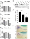
Bicc1 regulates Pkd2. (A-C) qPCR analysis for mouse Pkd1, Pkd2 and Pkhd1 mRNA levels in Bicc1+/+ (dark gray) and Bicc1−/− (light gray) littermates. The averages and s.d. from six kidney pairs at E15.5 and four pairs at E18.5 are shown (*, P<0.05, Student's t-test). (D) Western blot analysis comparing Pkd2 protein levels in E15.5 kidneys from two Bicc1 mouse litters (#55 and #65) of the indicated genotypes using the Pkd2-specific antibody from Santa Cruz. Actin served as a loading control. (E) Quantification of multiple Pkd2 western blot analyses comparing several mouse litters at E15.5 and normalized to actin. Average values and s.d. are indicated (*, P<0.05, Student's t-test). (F) Whole-mount in situ hybridization for Pkd2 mRNA on uninjected and xBicC-MO1+2-injected Xenopus embryos at stage 39.
Fig. 4.
Fig. 4.
Bicc1 is epistatic to Pkd2. (A-A″) Analysis of the expression of nbc-1 in the Xenopus late distal tubule at stage 39 by whole-mount in situ hybridization of uninjected control embryos, and embryos radially injected with xBicC-MO1+2 in the presence or absence of a single injection of 2 ng pkd2 mRNA. (B) Quantification of the expression of nbc-1 in the late distal tubule from the experiments shown in A-A″. Black, bilateral expression; white, no expression; gray, unilateral expression rescued by co-injected mRNA. The number of embryos analyzed is indicated. (C-D) Reciprocal experiments to those in A-B using Xenopus embryos injected with either Pkd2-MO alone or together with pkd2-myc or bicc1 mRNA. Co-injection with pkd2-myc rescued nbc-1 expression, whereas co-injection with bicc1 did not. (E) Flow diagram outlining the proposed mechanism of Bicc1 activity.
Fig. 5.
Fig. 5.
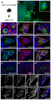
Subcellular localization of Bicc1. (A,B) mRNA encoding a Xenopus Bicc1-GFP fusion protein (xBicC-GFP) was injected into the animal region of Xenopus embryos and analyzed at gastrula stage by immunofluorescence microscopy. Inset is a magnified view of a single cell (arrow). (C-H″) HEK293T cells were transfected with plasmids encoding xBicC-GFP (C-G″) or a mouse Bicc1-GFP fusion protein (mBicC-GFP; H-H″) and processed for immunofluorescence with antibodies against GM130 (C), calregulin (D), Lamp2 (E), GW182 (F-F″) and HEDLs (G-H″), using red fluorescent secondary antibodies. Nuclei were counterstained with DAPI (blue). (I-J‴) HeLa cells were transfected with pCS2-xBicC-GFP and were either left untreated (I-I‴) or treated with 20 μM clotrimazole (J-J‴). Stress granule formation was visualized with antibodies against Eif4E (red) and Eif3η (blue). Note that even in the untreated cells, xBicC-GFP is partially colocalized with Eif4E, which is a marker for P-bodies and stress granules.
Fig. 6.
Fig. 6.
Post-transcriptional regulation by Bicc1. (A) qPCR analysis of the reduction in Pkd2 mRNA in the kidneys of Bicc1+/+ and Bicc1−/− mouse littermates, comparing total and capped mRNA at E15.5 and E18.5. (B) RL-PCR to determine the length of the poly(A) tail of Pkd2 mRNA, comparing kidneys of Bicc1+/+ and Bicc1−/− littermates at E15.5. As a control for the specificity of the reaction, oligo(dT) and RNase H were added to remove the poly(A) tail before the RL-PCR, which collapsed the smear seen in the gel into a distinct band. (C) Schematic representation of Pkd2 constructs [Pkd2(long_UTR), Pkd2(short_UTR), Pkd2(no_UTR), Pkd2(miR-17mut)]. Predicted conserved miRNA binding sites are indicated. (D,E) Western blot of Pkd2 expression in HEK293T cells transfected with the four Pkd2 constructs together with an empty vector control, Xenopus bicc1 or a bicc1 construct lacking the RNA-binding KH domains (xBicCΔKH). Equal loading was confirmed by actin. (F-F″) Quantification of three independent western blots showing mean values and s.d. (*, P<0.05). (G,G′) Xenopus embryos were injected with an mRNA containing the lacZ gene fused to the short or long 3′UTR of Pkd2 in the presence or absence of a miR-17 duplex and stained for lacZ expression at stage 10 (G). Multiple experiments were quantified and the number of embryos analyzed is indicated (G′). White, no lacZ staining; black, strong lacZ staining; gray, reduced lacZ staining. (H) Luciferase reporter assay of mouse embryonic fibroblasts (MEFs) transfected with pmirGLO-Pkd2-3′UTR and pmirGLO-Pkd2-3′UTR-mut in the presence of pCS2, pCS2-miR-17 or pCS2-miR-30a-5p. Values were corrected for the expression of Renilla luciferase and calculated as fold change compared with the pCS2 control. Multiple independent experiments were averaged and the s.d. is indicated (P<0.05).
Fig. 7.
Fig. 7.
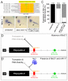
Cross-talk between Bicc1 and the miR-17 miRNA family. (A) Alignment of the Xenopus miR-17 family members. Mature forms are highlighted in yellow. The sequence targeted by the miR-17 antisense MO (miR-17-MO) is indicated by the black line. The nucleotides shared between miR-17-MO and the individual members are indicated in red. (B-B″) Analysis of the expression of nbc-1 by whole-mount in situ hybridization of uninjected control embryos, embryos injected with xBicC-MO1+2 alone or with miR-17-MO. Arrowheads indicate the expression of nbc-1 in the Xenopus late distal tubule. Note that this expression domain is rescued upon co-injection of the two antisense MOs. (C) Quantification of the experiments shown in B-B″. Black, bilateral expression; white, reduced or no expression; gray, unilateral, rescued expression in the late distal tubule. (D,D′) Models for the post-transcriptional regulation of Pkd2 mRNA by the miR-17 family in the absence or presence of Bicc1.

Similar articles

Cited by

References

    1. Agrawal R., Tran U., Wessely O. (2009). The miR-30 miRNA family regulates Xenopus pronephros development and targets the transcription factor Xlim1/Lhx1. Development 136, 3927-3936 - PMC - PubMed
    1. Anderson P., Kedersha N. (2008). Stress granules: the Tao of RNA triage. Trends Biochem. Sci. 33, 141-150 - PubMed
    1. Blum M., Andre P., Muders K., Schweickert A., Fischer A., Bitzer E., Bogusch S., Beyer T., van Straaten H. W., Viebahn C. (2007). Ciliation and gene expression distinguish between node and posterior notochord in the mammalian embryo. Differentiation 75, 133-146 - PubMed
    1. Burn T. C., Connors T. D., Dackowski W. R., Petry L. R., Van Raay T. J., Millholland J. M., Venet M., Miller G., Hakim R. M., Landes G. M., et al. (1995). Analysis of the genomic sequence for the autosomal dominant polycystic kidney disease (PKD1) gene predicts the presence of a leucine-rich repeat. The American PKD1 Consortium (APKD1 Consortium). Hum. Mol. Genet. 4, 575-582 - PubMed
    1. Charlesworth A., Cox L. L., MacNicol A. M. (2004). Cytoplasmic polyadenylation element (CPE)- and CPE-binding protein (CPEB)-independent mechanisms regulate early class maternal mRNA translational activation in Xenopus oocytes. J. Biol. Chem. 279, 17650-17659 - PMC - PubMed

Publication types

MeSH terms