. 2010 Apr;137(7):1205-13.
doi: 10.1242/dev.041251.

PHOX2A regulation of oculomotor complex nucleogenesis

Affiliations

PHOX2A regulation of oculomotor complex nucleogenesis

Khaleda B Hasan et al. Development. 2010 Apr.

Abstract

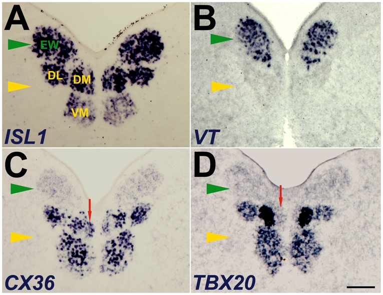
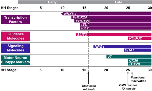
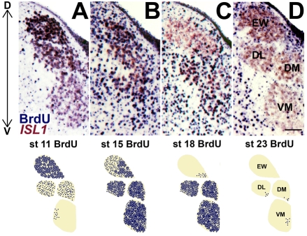
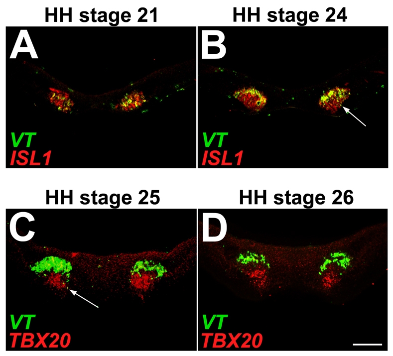
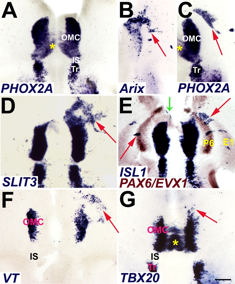
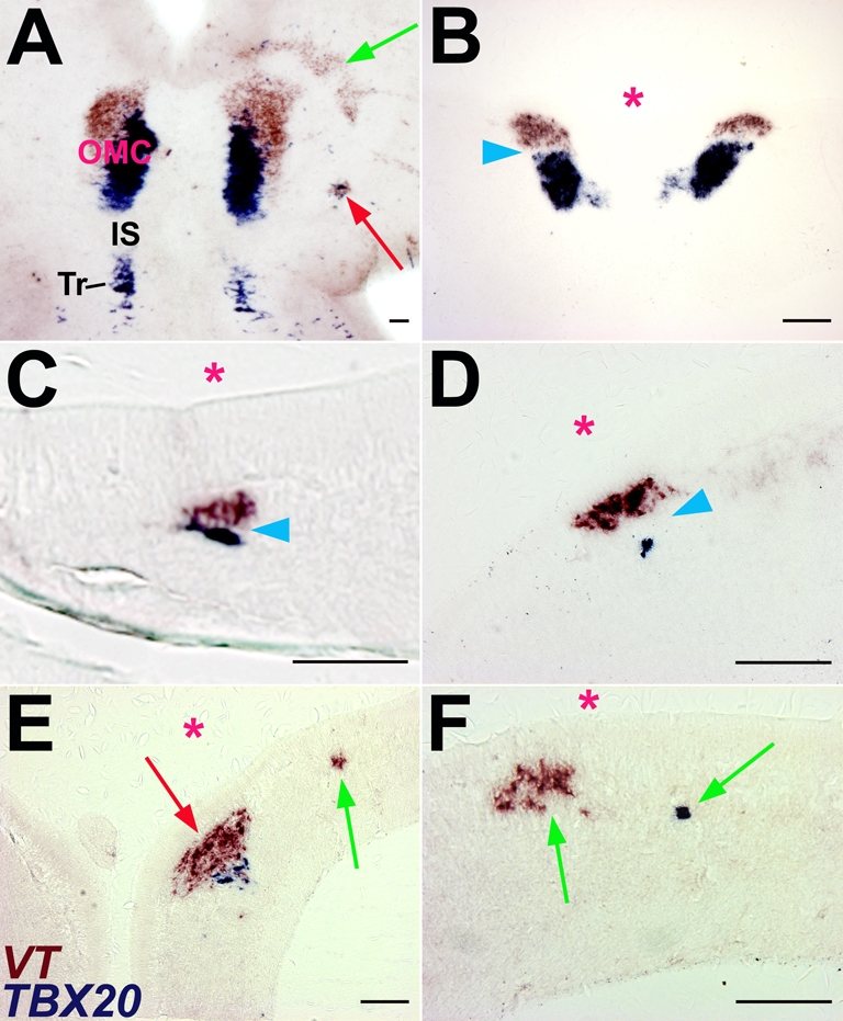
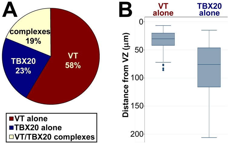
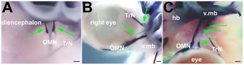
Brain nuclei are spatially organized collections of neurons that share functional properties. Despite being central to vertebrate brain circuitry, little is known about how nuclei are generated during development. We have chosen the chick midbrain oculomotor complex (OMC) as a model with which to study the developmental mechanisms of nucleogenesis. The chick OMC comprises two distinct cell groups: a dorsal Edinger-Westphal nucleus of visceral oculomotor neurons and a ventral nucleus of somatic oculomotor neurons. Genetic studies in mice and humans have established that the homeobox transcription factor gene PHOX2A is required for midbrain motoneuron development. We probed, in forced expression experiments, the capacity of PHOX2A to generate a spatially organized midbrain OMC. We found that exogenous Phox2a delivery to embryonic chick midbrain can drive a complete OMC molecular program, including the production of visceral and somatic motoneurons. Phox2a overexpression was also able to generate ectopic motor nerves. The exit points of such auxiliary nerves were invested with ectopic boundary cap cells and, in four examples, the ectopic nerves were seen to innervate extraocular muscle directly. Finally, Phox2a delivery was able to direct ectopic visceral and somatic motoneurons to their correct native spatial positions, with visceral motoneurons settling close to the ventricular surface and somatic motoneurons migrating deeper into the midbrain. These findings establish that in midbrain, a single transcription factor can both specify motoneuron cell fates and orchestrate the construction of a spatially organized motoneuron nuclear complex.


Figures

Fig. 1.
Fig. 1.
Gene expression markers distinguish the somatic and visceral motoneuron subnuclei of the chick oculomotor complex (OMC). Coronal sections of E12 chick midbrain processed for in situ hybridization to identify the somatic motoneurons of the oculomotor nucleus (yellow arrowhead) and the visceral motoneurons of the Edinger-Westphal nucleus (EW, green arrowhead). (A) The motoneuron marker ISL1 identifies all OMC motoneurons. (B) Gene expression for the neuropeptide VT selectively labels the visceral motoneurons. (C,D) The gap junction subunit gene CX36 (C) and the T-box transcription factor TBX20 (D) are expressed by the motoneurons of all somatic subnuclei. Within the DM subnucleus, TBX20 is strongly expressed in its lateral division, but is absent from its medial division (red arrow). DL, dorsolateral; DM, dorsomedial; VM, ventromedial. Scale bar: 0.2 mm.
Fig. 2.
Fig. 2.
Schedule of OMC development. Gene expression onset and timing of key events in chick oculomotor nerve (OMN) development, illustrated with reference to Hamburger-Hamilton (HH) stages (see text for details). Expression of the ligand-encoding SLIT3 and neuregulin-1 (NRG1) genes and of the SLIT receptor ROBO2 was detected by st 13+, st 17 and st 25, respectively. Timing of functional innervation is from Martinov et al. (Martinov et al., 2004). IO, inferior oblique.
Fig. 3.
Fig. 3.
Inside-out gradient of OMC neurogenesis. Chick midbrains were processed at E12 for BrdU immunohistochemistry (blue) and ISL1 gene expression (brown/pink) after BrdU delivery to st 8-23 embryos. Coronal sections (top) and corresponding BrdU chartings (bottom) illustrate the dorsal-to-ventral (D↔V) progression of OMC neurogenesis. Panels illustrate the left OMC, with the midline to the right. OMC subnuclei are identified in D. (A) Heavy BrdU labeling in the EW nucleus indicates that neurogenesis of visceral oculomotor neurons precedes that of somatic oculomotor neurons. (B) Extensive BrdU labeling is seen throughout the OMC. (C,D) Neurogenesis is nearly complete for the EW nucleus by st 18 (C) and for the somatic motoneurons by st 23 (D). Scale bar: 0.1 mm.
Fig. 4.
Fig. 4.
OMC motoneuron subtype segregation at the end of OMC neurogenesis. (A-D) Coronal sections of chick midbrains of the indicated stages processed for two-color fluorescent in situ hybridization. The ventricle is to the top and the pial surface is to the bottom. For st 21 (A) and st 24 (B), before TBX20 gene expression onset, VT (green) was employed to label visceral motoneurons and ISL1 (red) served to identify all OMC neurons. Because ISL1 co-labels VT-expressing cells, many visceral motoneurons appear yellow. St 25 (C) and st 26 (D) midbrains probed for VT (green) to mark visceral motoneurons and TBX20 (red) to label somatic motoneurons. At st 21, somatic motor production and migration is still underway and OMC subtypes appear intermixed (A). Once somatic motoneuron production has ended (B,C), few visceral motoneurons are found in the prospective somatic motoneuron territory (arrows). By st 26, the motoneuron classes are fully segregated, with a gap between the populations (D). Scale bar: 0.1 mm.
Fig. 5.
Fig. 5.
Phox2a induction of OMC gene expression markers. (A) Open-book preparation of E6 chick brainstem whole-mount demonstrates native PHOX2A gene expression in the midbrain OMC and the rostral hindbrain trochlear nucleus (Tr). Rostral is to the top, and the ventricular surface faces the viewer. (B-G) Expression plasmids for rat Phox2a (Arix) were electroporated into E2 chick ventral midbrain and embryos harvested for whole-mount in situ hybridization at E5 (D) and E6 (B,C,E-G). (B) A typical pattern of plasmid delivery by electroporation illustrated in the right midbrain by transgene expression (arrow). (C) Phox2a misexpression elicits induction of endogenous chick PHOX2A (arrow). (D) Phox2a forced expression induces other OMC markers, including the axon guidance molecule SLIT3 (arrow). (E) Lateral arcuate markers PAX6 (P6) and EVX1 (E1) (brown) illustrate the capacity of Phox2a to induce ISL1 (blue) throughout the ventral midbrain (red arrows) and rostrally in caudoventral diencephalon (green arrow). (F,G) Phox2a misexpression induces molecular markers (arrows) of visceral (VT, F) and somatic oculomotor neurons (TBX20, G). Asterisks (A,C,G) mark the contralateral migration of the prospective superior rectus somatic motoneurons (see Chilton and Guthrie, 2004). IS, isthmus. Scale bar: 0.2 mm.
Fig. 6.
Fig. 6.
Phox2a induction of mini-OMCs. Chick ventral midbrains electroporated with rat Phox2a plasmids at E2 and collected at E6. Tissue was processed to demonstrate visceral (VT, brown) and somatic (TBX20, blue) motoneurons. (A) Two-color in situ hybridization whole-mount, oriented and labeled as in Fig. 5A, demonstrates the ectopic induction of low-density fields of VT-expressing cells (green arrow) and clusters containing both VT-expressing and TBX20-expressing cells (red arrow). In this sample, the overlap of the VT-expressing and TBX20-expressing cells in the native OMC is not seen because of the density of TBX20 labeling, and the extent of the VT-positive territory is exaggerated by whole-mount flattening. (B) Entopic arrangement of the E6 OMC demonstrated in coronal cross-section, with the ventricle to the top (asterisk). VT-expressing visceral motoneurons sit closer to the ventricle, whereas TBX20-expressing somatic motoneurons lie deeper, closer to the pial surface, with a gap between the subtypes (arrowhead). (C-F) Coronal cross-sections through the ventral midbrain show the segregation of ectopic motoneuron classes along the ventricular-pial axis (ventricle marked by asterisk). A clear gap is often (C,D, arrowheads), but not always (E, red arrow), seen between the two classes of ectopic motoneurons. Forced Phox2a expression also produced isolated VT-expressing (E,F) and TBX20-expressing (F) cells in low-density fields (green arrows). Scale bars: 0.1 mm.
Fig. 7.
Fig. 7.
Ectopic visceral and somatic motoneurons have intrinsic migration programs. (A) Statistics on ectopic VT and TBX20 gene expression in four chick midbrains studied in serial section. In this sample, ectopic VT-expressing cells appeared more frequently (58%) without adjacent TBX20-expressing cells than in mini-OMCs (19%). Isolated Phox2a-induced TBX20-expressing cells accounted for 23% of the sample. (B) For each occurrence of cells expressing only VT or TBX20, the distance from the ventricular zone along the ventricular-pial axis was measured. These distances were compared using the two-sample Wilcoxon rank sum test. The horizontal line in the box represents the median value, and the edges of the box are the first and third quartiles. The lines extending from the edges of the box end at the last value within 1.5 times the interquartile range (third minus first quartile) from the edge of the box. Values beyond this distance are considered outliers and are plotted individually. Distances from the ventricular zone were statistically significantly greater for TBX20-expressing than for VT-expressing cells (see text).
Fig. 8.
Fig. 8.
Phox2a misexpression in ventral midbrain drives the production of ectopic cranial nervelets. Co-electroporation of alkaline phosphatase-expressing vector with rat Phox2a plasmid into ventral chick midbrain at E2 demonstrates ectopic axon outgrowth 4 days later, at harvesting. Mesenchyme was partially dissected away to show alkaline phosphatase-labeled nerves. Arrows point to ectopic axons. (A) Lateral view of an E6 embryo illustrating ectopic rootlets projecting out of ventral midbrain and ending in the subjacent mesenchyme. (B) Some ectopic axons join the oculomotor (III) and trochlear (IV) cranial nerves. (C) An ectopic nerve is seen to travel directly to the orbit, reaching the superior rectus muscle and bypassing nearby cranial nerves altogether. hb, hindbrain; OMN, oculomotor nerve; TrN, trochlear nerve; v.mb, ventral midbrain. Scale bars: 0.2 mm.
Fig. 9.
Fig. 9.
Phox2a-induced motoneurons organize boundary cap cells (BCCs) at ectopic motor nerve exits. (A-D) Rat Phox2a was delivered to the right side of E2 chick ventral midbrain. Heads were harvested 1-3 days later and processed for whole-mount in situ hybridization for the BCC markers CDH7 (A-C) and EGR2 (D). (A,D) Lateral views of right side of E3 (A) and E4 (D) heads. CDH7 (A) and EGR2 (D) labeling identifies BCCs at the native oculomotor nerve exit points (green arrows) and ectopic motor exit points (red arrows). (B,C) E5 Phox2a-electroporated heads probed for CDH7 expression and studied in cross-section. Sections cut transverse (B) and parallel (C) to the third cranial nerve demonstrate CDH7 expression at native (green arrows) and ectopic (red arrows) motor exit points. Ventricle in C is indicated by an asterisk. dien, diencephalon; fb, forebrain; hb, hindbrain; v.mb, ventral midbrain. Scale bars: 0.1 mm.

Similar articles

Cited by

References

    1. Agarwala S., Ragsdale C. W. (2002). A role for midbrain arcs in nucleogenesis. Development 129, 5779-5788 - PubMed
    1. Agarwala S., Ragsdale C. W. (2009). Midbrain patterning. In Encyclopedia of Neuroscience, Vol. 5 (ed. Squire L. R.), pp. 879-888 Amsterdam: Elsevier;
    1. Agarwala S., Sanders T. A., Ragsdale C. W. (2001). Sonic Hedgehog control of size and shape in midbrain pattern formation. Science 291, 2147-2150 - PubMed
    1. Bayly R. D., Ngo M., Aglyamova G. V., Agarwala S. (2007). Regulation of ventral midbrain patterning by Hedgehog signaling. Development 134, 2115-2124 - PubMed
    1. Bosley T. M., Oystreck D. T., Robertson R. L., al Awad A., Abu-Amero K., Engle E. C. (2006). Neurological features of congenital fibrosis of the extraocular muscles type 2 with mutations in PHOX2A. Brain 129, 2363-2374 - PubMed

Publication types