. 2010 Jul;38(12):3984-98.
doi: 10.1093/nar/gkq144. Epub 2010 Mar 9.

Telomeric protein TRF2 protects Holliday junctions with telomeric arms from displacement by the Werner syndrome helicase

Affiliations

Telomeric protein TRF2 protects Holliday junctions with telomeric arms from displacement by the Werner syndrome helicase

Gerald J Nora et al. Nucleic Acids Res. 2010 Jul.

Abstract

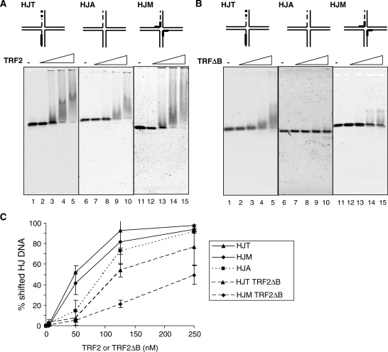
WRN protein loss causes Werner syndrome (WS), which is characterized by premature aging as well as genomic and telomeric instability. WRN prevents telomere loss, but the telomeric protein complex must regulate WRN activities to prevent aberrant telomere processing. Telomere-binding TRF2 protein inhibits telomere t-loop deletion by blocking Holliday junction (HJ) resolvase cleavage activity, but whether TRF2 also modulates HJ displacement at t-loops is unknown. In this study, we used multiplex fluorophore imaging to track the fate of individual strands of HJ substrates. We report the novel finding that TRF2 inhibits WRN helicase strand displacement of HJs with telomeric repeats in duplex arms, but unwinding of HJs with a telomeric center or lacking telomeric sequence is unaffected. These data, together with results using TRF2 fragments and TRF2 HJ binding assays, indicate that both the TRF2 B- and Myb domains are required to inhibit WRN HJ activity. We propose a novel model whereby simultaneous binding of the TRF2 B-domain to the HJ core and the Myb domain to telomeric arms promote and stabilize HJs in a stacked arm conformation that is unfavorable for unwinding. Our biochemical study provides a mechanistic basis for the cellular findings that TRF2 regulates WRN activity at telomeres.


Figures

Figure 1.
Figure 1.
WRN helicase and exonuclease simultaneously process HJ substrates with a mobile core. (A) HJA (2 nM) was incubated with 24 nM WRN in standard reaction buffer at 37°C. Aliquots were terminated at various times from 0.5 to 60 min in 2-fold increments (lanes 2–10). Reactions were run on a 12% native polyacrylamide gel and were visualized with a Typhoon Imager in the TAMRA (blue), Cy5 (green) and Alexa 488 (red) channels. Colors assigned by Imagequant software. The exonuclease degraded products are indicated as (a) = exo-fork and (b) = exo-ssDNA. (B) Schematic of WRN strand displacement and exonuclease products. T = TAMRA, C = Cy5 and A = Alexa488 labeled oligonucleotides. The 3′ end of the exonuclease vulnerable T-oligo is highlight as a dotted line. (C) The percent of displaced T-oligo products from the reactions in (A) were quantitated as described in ‘Materials and Methods’ section and plotted against time. Intact fork, solid black line and squares; exonuclease-degraded fork, dashed black line and triangles; intact ssDNA, solid gray line and squares; exonuclease-degraded ssDNA, dashed gray line and triangles. (D) Substrate and product distribution for each labeled oligonucleotide in the HJ construct after 1 h reaction. The percent of T-, A- and C-oligos present in the HJ substrate and each intermediate and product were quantitated as a function of total oligonucleotide as described in ‘Materials and Methods’. Values represent the mean and standard deviation of two or three separate experiments.
Figure 2.
Figure 2.
A biotin–streptavidin complex on the translocating strand of a forked duplex inhibits WRN unwinding activity. Reactions contained a 31-bp forked duplex (0.5 nM) with a biotinylated nucleotide (inverted trapezoid) on the WRN translocating (A) or nontranslocating strand (B). Black box denotes streptavidin. The forks were constructed by annealing oligonucleotides bio-1 and 4 (A) or bio-1 and 2 (B) (Supplementary Table S1). T = TAMRA; A = Alexa488. The duplex length (bp) on either side of the biotinylated nucleotide is shown. The substrate was pre-incubated with either 0 (lanes 1 and 2) or 30 nM (lanes 3 and 4) streptavidin in standard reaction buffer prior to adding 3.4 nM X-WRN and 12 nM RPA. Reactions were for 20 min at 37°C, and were run on an 8% native polyacrylamide gel for 2.5 h and visualized with a Typhoon imager. A streptavidin-bound T-oligo was loaded (lane 6) as a marker and the boiled substrate lane is indicated with a triangle (lane 5). The gel scans show the TAMRA emission channel. (C) Quantitation of the ssDNA product. The percent of ssDNA T-oligo product was quantitated as described in ‘Materials and Methods’ section. Values represent the mean and SEM. from two independent experiments.
Figure 3.
Figure 3.
A biotin–streptavidin complex at the HJ core impedes WRN helicase activity. (A) The HJbio-center construct has a 12-nt homologous core that permits branch migration. The center construct (II) shows the HJ in a symmetric conformation with 25-bp arms. The left and right constructs (I and III) show the possible extreme conformations. T = TAMRA; C = Cy5; A = Alexa488; inverted trapezoid denotes a biotin moiety. Black box denotes a streptavidin. The predicated possible directions of unwinding are shown for each conformation; white arrow denotes unwinding along the horizontal axis; black arrow denotes unwinding along the vertical axis. (B and C) The HJbio-center substrate (0.5 nM) was pre-incubated without (lanes 1 and 2) or with (lanes 4 and 5) 30 nM streptavidin under standard reaction conditions prior the addition of 15 nM X-WRN and 45 nM RPA. The reactions were conducted for 1 h at 37°C. The products were run on an 8% native acrylamide gel for 2.5 h and visualized with a Typhoon Imager. Black triangle denotes boiled substrate lane. The TAMRA and Alexa488 emission channels are shown in (B) and (C), respectively. (D) Quantitation of ssDNA reaction products. The percent of T-oligo (gray bars) and A-oligo (white bars) detected as ssDNA product was quantitated as described in ‘Materials and Methods’ section. The values represent the mean and standard deviation from two to four independent experiments. (E) Schematic of the three-way product species that are generated upon addition of streptavidin.
Figure 4.
Figure 4.
A biotin–streptavidin complex at the 3′-end of an HJ arm does not alter WRN helicase activity. The HJbio-end substrate has a biotin tag at the 3′-end of the A-oligo. T = TAMRA; C = Cy5; A = Alexa488; inverted trapezoid denotes a biotin moiety. Black box denotes streptavidin. (A) The HJbio-end (0.5 nM) substrate was pre-incubated without (lanes 2–4) or with (lanes 6 and 7) 30 nM streptavidin under standard reaction conditions prior the addition of 15 nM X-WRN and 45 nM RPA. The reactions were conducted for 1 h at 37°C. The products were run on an 8% native acrylamide gel for 2.5 h and visualized with a Typhoon Imager. Black triangle denotes boiled lane. M denotes an A-oligo ssDNA marker bound by streptavidin. The Alexa488 emission channel is shown. (B) Quantitation of ssDNA reaction products. The percent A-oligo detected as ssDNA product was quantitated as described in ‘Materials and Methods’ section. The values represent the mean and standard deviation from three independent experiments.
Figure 5.
Figure 5.
TRF2 inhibits WRN unwinding of HJ substrates with telomeric arms. A schematic of the telomeric arm (HJT) and non-telomeric HJA constructs are shown in (A–C). Thick black lines denote (TTAGGG)3 repeats. T = TAMRA; C = Cy5; A = Alexa488. The 3′-end of the exonuclease-vulnerable T-oligo is highlighted as a dotted line. Predicted TRF2 binding sites are indicated with the circle (B-domain) and the ellipse (Myb domain). The substrates (0.5 nM) HJT (A) or HJA (B) were pre-incubated with 0, 0.5, 5, 12.5 or 25 nM TRF2 [(A), lanes 2–6, respectively] and 0, 0.5, 5 or 25 nM TRF2 [(B), lanes 1–4] in standard reaction buffer. The reactions were initiated by the addition of 19 nM WRN and reacted for 1 h at 37°C. The reactions were run on 8% native gels for 1.5 h and visualized with a Typhoon Imager. Scans of the Cy5 emission channel are shown in (A) and (B). Triangle indicates boiled substrate; M denotes a marker lane; arrows denote a triple-stranded species. (C) TRF2ΔB does not alter WRN unwinding of the telomeric HJ. The HJT (0.5 nM) substrate was pre-incubated with 0, 0.5, 5 or 25 nM TRF2ΔB (lanes 2–5) prior to the addition of 19 nM WRN. Reactions were for 1 h at 37°C, and were run on an 8% native gel. The scan from the Cy5 emission channel is shown. (D) Quantitation of HJ unwinding. The percent of C-oligo present in the HJ substrate was quantitated as a function of total C-oligo DNA in the reaction as described in ‘Materials and Methods’ section and plotted against TRF2 or TRF2ΔB concentration. The values represent the mean and standard deviation from at least three independent experiments.
Figure 6.
Figure 6.
TRF2 fails to inhibit WRN helicase or exonuclease activities on an HJ with telomeric repeats confined to the core. (A) The HJM substrate has a 12-nt homologous core composed of two (TTAGGG)2 repeats, denoted by a thick black line. Shown is the conformation with a fully branch migrated core. The predicted TRF2 binding sites are indicated with the circle (B-domain) and the ellipse (Myb domain). C = Cy5 fluorophore. The 3′-end of the exonuclease vulnerable C-oligo is highlighted as a dotted line. (B and C) The HJM substrate (0.5 nM) was pre-incubated with either 0, 0.5, 5, 12.5 or 25 nM TRF2 (lanes 3–7) and reacted under standard conditions for 1 h with 25 nM WRN (B) or 40 nM X-WRN (C), respectively. The reactions were run on 8% native gels for 1.5 h and visualized with a Typhoon Imager. The scan from the Cy5 emission channel is shown. Triangle indicates boiled substrate. (D) Quantitation of HJ unwinding. The percent of C-oligo in the HJM displacement products were quantitated as a function of total C-oligo DNA as described in ‘Materials and Methods’ section and plotted against the TRF2 concentration. The values represent the mean and standard deviation from three separate experiments.
Figure 7.
Figure 7.
The RAP1/TRF2 complex inhibits WRN unwinding of HJ substrates with telomeric arms similar to TRF2 alone. (A) Schematic of the HJT construct is shown. Thick black lines denote (TTAGGG)3 repeats. T = TAMRA; C = Cy5; A = Alexa488. The 3′-end of the exonuclease vulnerable T-oligo is highlight as a dotted line. Predicted TRF2 binding sites are indicated with the circle (B-domain) and the ellipse (Myb domain). (B) The substrate was pre-incubated with 25 nM TRF2 and 0, 12.5, 25 or 50 nM RAP1 (lanes 2–5) in standard reaction buffer. The reactions were initiated by the addition of 19 nM WRN (lanes 1–6) and reacted for 1 h at 37°C. Lane 6 contained 50 nM RAP1 and 19 nM WRN. The reactions were run on 8% native gels and visualized with a Typhoon Imager. Scans of the Cy5 emission channel are shown. Triangle indicates boiled substrate; arrows denote a triple-stranded species. (C) Quantitation of HJ unwinding. The percent of C-oligo present in the HJ products was quantitated as a function of total C-oligo DNA as described in ‘Materials and Methods’ section and plotted against RAP1 concentration.
Figure 8.
Figure 8.
TRF2 and TRF2ΔB exhibit increased binding to telomeric HJ compared to non-telomeric HJ constructs. The substrates (5 nM) telomeric arm HJT (lanes 1–5), non-telomeric HJA (lanes 6–10), and telomeric core HJM (lanes 11–15) were reacted with either 0, 5, 50, 125 or 250 nM TRF2 (A) or TRF2ΔB (B), respectively, in standard reaction buffer supplemented with 5% glycerol for 20 min at 24°C. The reactions were loaded on a 1% 0.5× TBE agarose gel and electrophoresed for 1 h at 140 V in 4°C. Gels were scanned on a Typhoon Imager and visualized in the Cy5 channel. (C) Quantitation of the binding reactions. The percent of bound substrate was calculated as a function of total DNA in the Cy5 channel and plotted against TRF2 or TRF2ΔB concentration. Values represent the mean and standard deviation from two or three independent reactions.

Similar articles

Cited by

References

    1. Kudlow BA, Kennedy BK, Monnat RJ., Jr Werner and Hutchinson-Gilford progeria syndromes: mechanistic basis of human progeroid diseases. Nat. Rev. Mol. Cell Biol. 2007;8:394–404. - PubMed
    1. Opresko PL. Telomere ResQue and preservation–roles for the Werner syndrome protein and other RecQ helicases. Mech. Ageing Dev. 2008;129:79–90. - PubMed
    1. Huang S, Li B, Gray MD, Oshima J, Mian IS, Campisi J. The premature ageing syndrome protein, WRN, is a 3′–>5′ exonuclease. Nat. Genet. 1998;20:114–116. - PMC - PubMed
    1. Chu WK, Hickson ID. RecQ helicases: multifunctional genome caretakers. Nat. Rev. Cancer. 2009;9:644–654. - PubMed
    1. Sidorova JM, Li N, Folch A, Monnat RJ., Jr The RecQ helicase WRN is required for normal replication fork progression after DNA damage or replication fork arrest. Cell Cycle. 2008;7:796–807. - PMC - PubMed

Publication types

MeSH terms