

Targeted inhibition of inducible nitric oxide synthase inhibits growth of human melanoma in vivo and synergizes with chemotherapy
- PMID: 20215556
- PMCID: PMC2858983
- DOI: 10.1158/1078-0432.CCR-09-3123
Targeted inhibition of inducible nitric oxide synthase inhibits growth of human melanoma in vivo and synergizes with chemotherapy
Abstract
Purpose: Aberrant expression of inflammatory molecules, such as inducible nitric oxide (NO) synthase (iNOS), has been linked to cancer, suggesting that their inhibition is a rational therapeutic approach. Whereas iNOS expression in melanoma and other cancers is associated with poor clinical prognosis, in vitro and in vivo studies suggest that iNOS and NO can have both protumor and antitumor effects. We tested the hypothesis that targeted iNOS inhibition would interfere with human melanoma growth and survival in vivo in a preclinical model.
Experimental design: We used an immunodeficient non-obese diabetic/severe combined immunodeficient xenograft model to test the susceptibility of two different human melanoma lines to the orally-given iNOS-selective small molecule antagonist N(6)-(1-iminoethyl)-l-lysine-dihydrochloride (L-nil) with and without cytotoxic cisplatin chemotherapy.
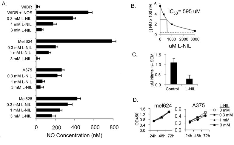
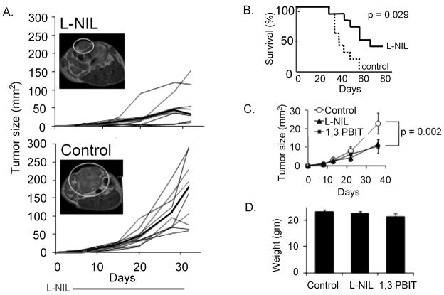
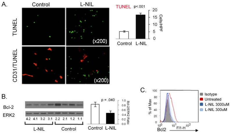
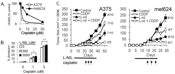
Results: L-nil significantly inhibited melanoma growth and extended the survival of tumor-bearing mice. L-nil treatment decreased the density of CD31+ microvessels and increased the number of apoptotic cells in tumor xenografts. Proteomic analysis of melanoma xenografts with reverse-phase protein array identified alterations in the expression of multiple cell signaling and survival genes after L-nil treatment. The canonical antiapoptotic protein Bcl-2 was downregulated in vivo and in vitro after L-nil treatment, which was associated with increased susceptibility to cisplatin-mediated tumor death. Consistent with this observation, combination therapy with L-nil plus cisplatin in vivo was more effective than either drug alone, without increased toxicity.
Conclusions: These data support the hypothesis that iNOS and iNOS-derived NO support tumor growth in vivo and provide convincing preclinical validation of targeted iNOS inhibition as therapy for solid tumors.
Figures


Similar articles
-
iNOS expression in CD4+ T cells limits Treg induction by repressing TGFβ1: combined iNOS inhibition and Treg depletion unmask endogenous antitumor immunity.Clin Cancer Res. 2014 Dec 15;20(24):6439-51. doi: 10.1158/1078-0432.CCR-13-3409. Epub 2014 Oct 2. Clin Cancer Res. 2014. PMID: 25278453
-
Chronic inhibition of inducible nitric oxide synthase ameliorates cardiovascular abnormalities in streptozotocin diabetic rats.Eur J Pharmacol. 2009 Jun 2;611(1-3):53-9. doi: 10.1016/j.ejphar.2009.03.061. Epub 2009 Apr 1. Eur J Pharmacol. 2009. PMID: 19344709
-
Role of inducible nitric oxide synthase in induction of RhoA expression in hearts from diabetic rats.Cardiovasc Res. 2008 Jul 15;79(2):322-30. doi: 10.1093/cvr/cvn095. Epub 2008 Apr 14. Cardiovasc Res. 2008. PMID: 18411229
-
Effect of inhibition of inducible nitric oxide synthase (iNOS) on the murine splenic immune response induced by Aggregatibacter (Actinobacillus) actinomycetemcomitans lipopolysaccharide.Eur J Oral Sci. 2008 Feb;116(1):31-6. doi: 10.1111/j.1600-0722.2007.00501.x. Eur J Oral Sci. 2008. PMID: 18186729
-
Cationic liposome-mediated nitric oxide synthase gene therapy enhances the antitumor effects of cisplatin in lung cancer.Int J Mol Med. 2013 Jan;31(1):33-42. doi: 10.3892/ijmm.2012.1171. Epub 2012 Nov 1. Int J Mol Med. 2013. PMID: 23128378
Cited by
-
Functional involvement of β3-adrenergic receptors in melanoma growth and vascularization.J Mol Med (Berl). 2013 Dec;91(12):1407-19. doi: 10.1007/s00109-013-1073-6. Epub 2013 Aug 2. J Mol Med (Berl). 2013. PMID: 23907236
-
Biochemistry of nitric oxide.Indian J Clin Biochem. 2011 Jan;26(1):3-17. doi: 10.1007/s12291-011-0108-4. Epub 2011 Feb 3. Indian J Clin Biochem. 2011. PMID: 22211007 Free PMC article.
-
Role of Endogenous Nitric Oxide in Hyperaggressiveness of Tumor Cells that Survive a Photodynamic Therapy Challenge.Crit Rev Oncog. 2016;21(5-6):353-363. doi: 10.1615/CritRevOncog.2017020909. Crit Rev Oncog. 2016. PMID: 29431083 Free PMC article.
-
Gr-1+CD11b+ cells facilitate Lewis lung cancer recurrence by enhancing neovasculature after local irradiation.Sci Rep. 2014 Apr 29;4:4833. doi: 10.1038/srep04833. Sci Rep. 2014. PMID: 24776637 Free PMC article.
-
Chemoprevention of melanoma.Adv Pharmacol. 2012;65:361-98. doi: 10.1016/B978-0-12-397927-8.00012-9. Adv Pharmacol. 2012. PMID: 22959032 Free PMC article. Review.
References
-
- Kundu JK, Surh Y. Inflammation: gearing the journey to cancer. Mutat Res. 2008;659:15–30. - PubMed
-
- Vakkala M, et al. Inducible nitric oxide synthase expression, apoptosis, and angiogenesis in in situ and invasive breast carcinomas. Clin Cancer Res. 2000;6:2408–2416. - PubMed
-
- Brennan PA, et al. Inducible nitric oxide synthase: Correlation with extracapsular spread and enhancement of tumor cell invasion in head and neck squamous cell carcinoma. Head Neck. 2008;30:208–14. - PubMed
Publication types
MeSH terms
Substances
Grants and funding
LinkOut - more resources
Full Text Sources
Other Literature Sources
Medical
