

Review
doi: 10.4161/rna.7.2.11207.
Epub 2010 Mar 14.
Spliceosomal snRNA modifications and their function
Affiliations
- PMID: 20215871
- PMCID: PMC4154345
- DOI: 10.4161/rna.7.2.11207
Item in Clipboard
Review
Spliceosomal snRNA modifications and their function
RNA Biol.
2010 Mar-Apr.
Abstract
Spliceosomal snRNAs are extensively 2'-O-methylated and pseudouridylated. The modified nucleotides are relatively highly conserved across species, and are often clustered in regions of functional importance in pre-mRNA splicing. Over the past decade, the study of the mechanisms and functions of spliceosomal snRNA modifications has intensified. Two independent mechanisms behind these modifications, RNA-independent (protein-only) and RNA-dependent (RNA-guided), have been discovered. The role of spliceosomal snRNA modifications in snRNP biogenesis and spliceosome assembly has also been verified.
Figures
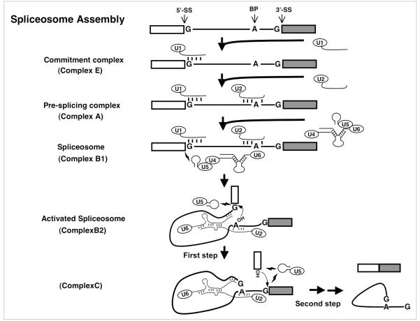
Major spliceosome assembly and catalysis of pre-mRNA splicing. The thick lines represent the intron and the boxes are exons. The 5' splice site (5'-SS), the 3' splice site (3'-SS) and the branch point adenosine (BP) are indicated in the pre-mRNA. The conserved residues at the 5' and 3' splice sites and the branch site are shown. The headed thin lines are snRNAs with their names in the ellipses. The short thick lines between RNA strands represent Watson-Crick base-pairing interactions. The lightning symbols depict non-Watson-Crick base-pairing interactions. The 2'-OH groups of branch point adenosine and the cut-off 5' exon are pictured in the activated spliceosome. The small arrows near those 2'-OH group indicate the nucleophilic chemical reactions also known as trans-esterification reactions.
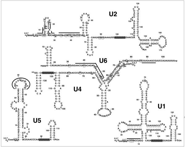
Pseudouridines and 2'-O-methylated residues in human spliceosomal snRNAs. Primary and secondary structures of human major spliceosomal snRNAs (U1, U2, U4, U5 and U6) are shown. Pseudouridines (Ψ) are surrounded by rectangles; 2'-O-methylations are circled. The thick lines indicate the nucleotides participating in RNA-RNA interactions or involved in catalysis during pre-mRNA splicing. The gray boxes highlight the Sm-binding sites. The 5' caps (2,2,7 trimethylated guanosine cap for U1, U2, U4, U5 and γ-methylated guanosine cap for U6) are also depicted.
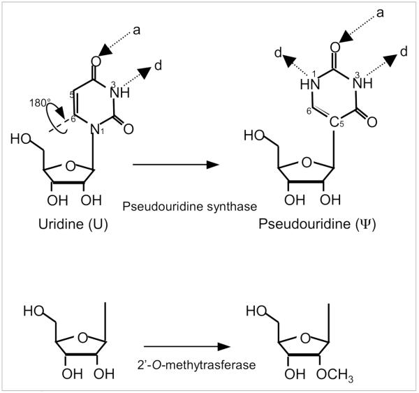
Schematic depiction of the two most abundant modified nucleotides in spliceosomal snRNA. (Top) Pseudouridine is a rotational isomer of uridine, in which the N-C glycosidic bond is broken to form an C-C bond. This results in the presence of an extra hydrogen bond donor (d), while the number of hydrogen bond acceptors (a) is unchanged. (Bottom) Schematic representation of a 2'-O-methylated ribose.
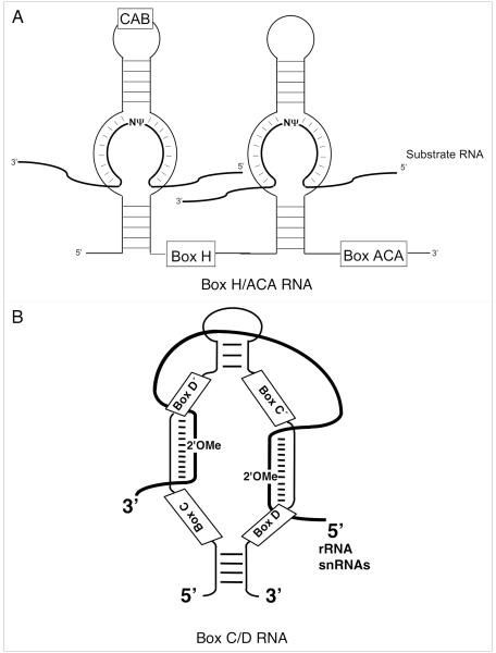
Schematic depiction of box H/ACA and C/D RNAs. (A) Secondary structure of a eukaryotic pseudouridylation guide box H/ACA RNA. The RNA adapts a Hairpin-Hinge-Hairpin-Tail structure. Present within the hinge region is the box H (5'-ANANNA-3'), the box ACA (5'-ACA-3') motif typically lies three nucleotides from the 3'-end of the RNA. A CAB box (5'-ugAG-3'), responsible for Cajal body localization, may be present in the apical loop of either hairpin. Pseudouridylation is targeted to substrate RNAs by complementary base-pairing interactions between the internal loop (pseudouridylation pocket) and nucleotides adjacent to the target uridine. The thick lines denote substrate RNAs. (B) Secondary structure of a box C/D RNA. Boxes C, D, C’ and D’ are shown. The 2'OMe represents the target 2'-O-methylation site that is always the fifth nucleotide from box D or D’. The thick line represents substrate RNA.
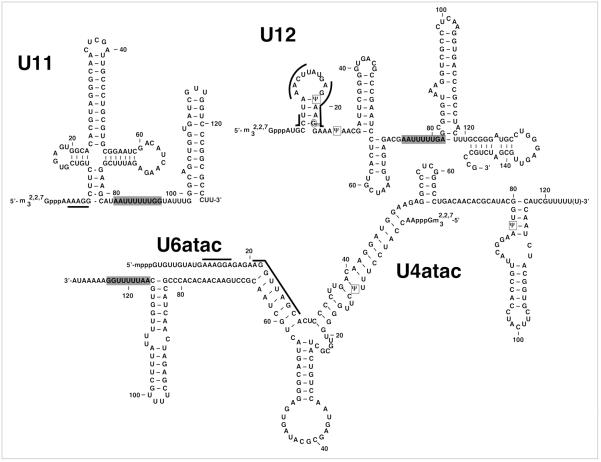
Shown are primary and secondary structures of human minor spliceosomal snRNAs, U11, U12, U4atac and U6atac. U5 snRNA is shared by both the major and minor spliceosomes. Pseudouridines (Ψ) are surrounded by rectangles; 2'-O-methylations are circled. Pseudouridines within U12 and U4atac are believed to function analogously to their homologous modifications within U2 and U4 snRNAs, respectively. The thick lines indicate the nucleotides participating in RNA-RNA interactions or involved in catalysis during pre-mRNA splicing. The gray boxes highlight the Sm-binding sites.
Similar articles
-
Functions and mechanisms of spliceosomal small nuclear RNA pseudouridylation.Wiley Interdiscip Rev RNA. 2011 Jul-Aug;2(4):571-81. doi: 10.1002/wrna.77. Epub 2011 Feb 18. Wiley Interdiscip Rev RNA. 2011. PMID: 21957045 Free PMC article. Review.
-
Pseudouridines in spliceosomal snRNAs.Protein Cell. 2011 Sep;2(9):712-25. doi: 10.1007/s13238-011-1087-1. Epub 2011 Oct 6. Protein Cell. 2011. PMID: 21976061 Free PMC article. Review.
-
Pseudouridine modification in Caenorhabditis elegans spliceosomal snRNAs: unique modifications are found in regions involved in snRNA-snRNA interactions.BMC Mol Biol. 2005 Oct 19;6:20. doi: 10.1186/1471-2199-6-20. BMC Mol Biol. 2005. PMID: 16236171 Free PMC article.
-
Localization of modified nucleotides in Schizosaccharomyces pombe spliceosomal small nuclear RNAs: modified nucleotides are clustered in functionally important regions.RNA. 1996 Sep;2(9):909-18. RNA. 1996. PMID: 8809017 Free PMC article.
-
Modified nucleotides at the 5' end of human U2 snRNA are required for spliceosomal E-complex formation.RNA. 2004 Dec;10(12):1925-33. doi: 10.1261/rna.7186504. Epub 2004 Nov 3. RNA. 2004. PMID: 15525712 Free PMC article.
Cited by
-
The epitranscriptome landscape of small noncoding RNAs in stem cells.Stem Cells. 2020 Oct 1;38(10):1216-1228. doi: 10.1002/stem.3233. Epub 2020 Jun 29. Stem Cells. 2020. PMID: 32598085 Free PMC article. Review.
-
Non-Coding RNAs in Breast Cancer: Intracellular and Intercellular Communication.Noncoding RNA. 2018 Dec 12;4(4):40. doi: 10.3390/ncrna4040040. Noncoding RNA. 2018. PMID: 30545127 Free PMC article. Review.
-
Site-Directed Spin Labeling of RNA with a Gem-Diethylisoindoline Spin Label: PELDOR, Relaxation, and Reduction Stability.Molecules. 2019 Dec 6;24(24):4482. doi: 10.3390/molecules24244482. Molecules. 2019. PMID: 31817785 Free PMC article.
-
The epitranscriptome of small non-coding RNAs.Noncoding RNA Res. 2021 Oct 26;6(4):167-173. doi: 10.1016/j.ncrna.2021.10.002. eCollection 2021 Dec. Noncoding RNA Res. 2021. PMID: 34820590 Free PMC article. Review.
-
Functions of RNA N6-methyladenosine modification in cancer progression.Mol Biol Rep. 2019 Apr;46(2):2567-2575. doi: 10.1007/s11033-019-04655-4. Epub 2019 Mar 25. Mol Biol Rep. 2019. PMID: 30911972 Review.
References
-
- Staley JP, Guthrie C. Mechanical devices of the spliceosome: motors, clocks, springs and things. Cell. 1998;92:315–26. - PubMed
-
- Yu YT, Scharl EC, Smith CM, Steitz JA. The RNA World. 2nd Cold Spring Harbor Laboratory Press; Cold Spring Harbor NY: 1999. pp. 487–524.
-
- Jurica MS, Moore MJ. Pre-mRNA splicing: awash in a sea of proteins. Mol Cell. 2003;12:5–14. - PubMed
-
- Valadkhan S. snRNAs as the catalysts of pre-mRNA splicing. Curr Opin Chem Biol. 2005;9:603–8. - PubMed
-
- Kramer A, Keller W, Appel B, Luhrmann R. The 5' terminus of the RNA moiety of U1 small nuclear ribonucleoprotein particles is required for the splicing of messenger RNA precursors. Cell. 1984;38:299–307. - PubMed
Publication types
MeSH terms
Substances
Grants and funding
LinkOut - more resources
Full Text Sources
Other Literature Sources
Molecular Biology Databases
Miscellaneous