

Correlation between morphologic features on spectral-domain optical coherence tomography and angiographic leakage patterns in macular edema
- PMID: 20216291
- PMCID: PMC2870721
- DOI: 10.1097/IAE.0b013e3181cd4803
Correlation between morphologic features on spectral-domain optical coherence tomography and angiographic leakage patterns in macular edema
Abstract
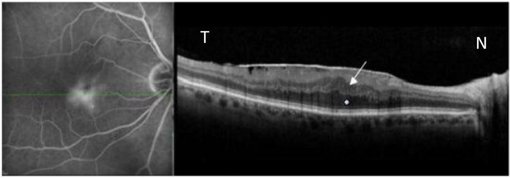
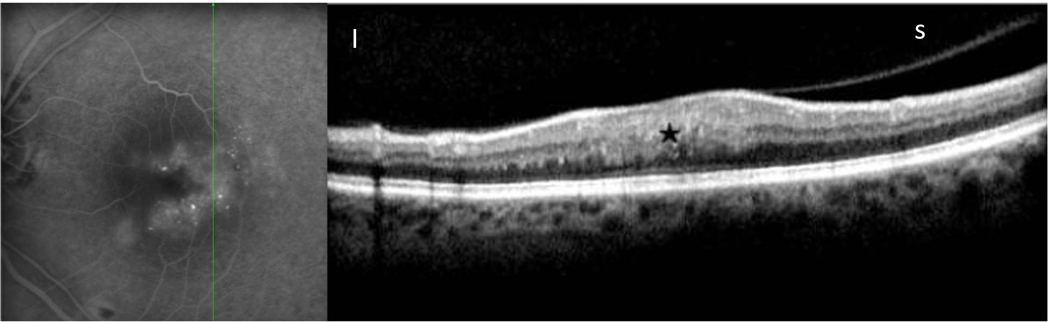
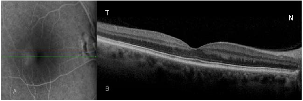
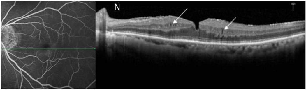
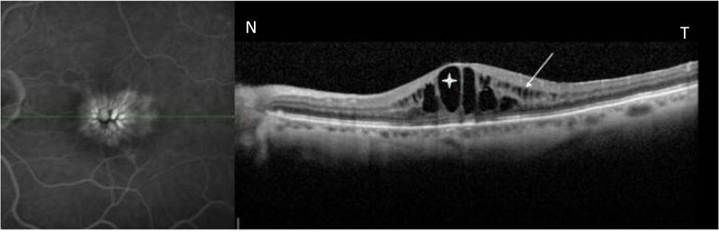
Purpose: The purpose of this study was to determine the morphologic patterns of angiographic macular edema using simultaneous colocalization of fluorescein angiography and spectral-domain optical coherence tomography (SD-OCT) images in diabetes, epiretinal membrane, uveitic and pseudophakic cystoid macular edema, and vein occlusion.
Methods: Eighty-seven consecutive patients (107 eyes) with macular edema from 5 different etiologies were imaged by simultaneous scanning laser ophthalmoscopy/OCT to study the morphologic patterns of edema on SD-OCT and then correlated/colocalized with the fluorescein angiographic patterns of leakage. Statistical analysis was done to analyze the differences in the morphologic OCT pattern by different diseases.
Results: Spectral-domain OCT characteristics of macular edema showed a significant difference across different diseases (P = 0.037). Cystic fluid pockets were found to be more commonly seen in patients with diabetic macular edema and retinal vein occlusions, whereas those cases with macular edema secondary to epiretinal membrane showed noncystic changes on OCT. Seventy of the 107 eyes had diffuse angiographic leakage, and the remaining 37 eyes had cystoid leakage on angiography. Of the 70 eyes with diffuse leakage, 24.28% showed microcysts on SD-OCT in the area of edema, and 70% eyes had diffuse thickening or distorted architecture without cyst. All 37 eyes with cystoid leakage showed cysts in the area of edema by SD-OCT. A total of 3.73% of eyes with fluorescein angiographic leakage had no abnormalities on SD-OCT.
Conclusion: Eyes with diabetic macular edema and retinal vein occlusions have a significantly higher incidence of cyst formation on SD-OCT. There was no correlation between visual acuity and cyst formation. Diffuse noncystoid angiographic macular edema may show microcysts on SD-OCT, but diffuse edema is more commonly associated with thickening or distortion of the retinal layers without cyst formation. Cystoid leakage on fluorescein angiography is always associated with cystic changes on SD-OCT.
Figures

References
-
- Marmor MF. Mechanisms of fluid accumulation in retinal edema. Doc Ophthalmol. 1999;97(3–4):239–249. - PubMed
-
- Schaudig U, Scholz F, Lerche RC, Richard G. Optical coherence tomography for macular edema. Classification, quantitative assessment, and rational usage in the clinical practice. Ophthalmologe. 2004;101(8):785–793. - PubMed
-
- Catier A, Tadayoni R, Paques M, et al. Characterization of macular edema from various etiologies by optical coherence tomography. Am J Ophthalmol. 2005;140(2):200–206. - PubMed
-
- Tran TH, de Smet MD, Bodaghi B, et al. Uveitic macular oedema: correlation between optical coherence tomography patterns with visual acuity and fluorescein angiography. Br J Ophthalmol. 2008;92(7):922–927. - PubMed
-
- Otani T, Kishi S, Maruyama Y. Patterns of diabetic macular edema with optical coherence tomography. Am J Ophthalmol. 1999;127(6):688–693. - PubMed
Publication types
MeSH terms
Grants and funding
LinkOut - more resources
Full Text Sources
Medical
