Changes in ocular aquaporin expression following optic nerve crush
- PMID: 20216911
- PMCID: PMC2831780
Changes in ocular aquaporin expression following optic nerve crush
Abstract
Purpose: Changes in the expression of water channels (aquaporins; AQP) have been reported in several diseases. However, such changes and mechanisms remain to be evaluated for retinal injury after optic nerve crush (ONC). This study was designed to analyze changes in the expression of AQP4 (water selective channel) and AQP9 (water and lactate channel) following ONC in the rat.
Methods: Rat retinal ganglion cells (RGCs) were retrogradely labeled by applying FluoroGold onto the left superior colliculus 1 week before ONC. Retinal injuries were induced by ONC unilaterally. Real-time PCR was used to measure changes in AQP4, AQP9, thy-1, Kir4.1 (K(+) channel), and beta-actin messages. Changes in AQP4, AQP9, Kir4.1, B cell lymphoma-x (bcl-xl), and glial fibrillary acidic protein (GFAP) expression were measured in total retinal extracts using western blotting.
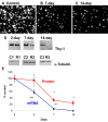
Results: The number of RGCs labeled retrogradely from the superior colliculus was 2,090+/-85 cells/mm(2) in rats without any treatment, which decreased to 1,091+/-78 (47% loss) and 497+/-87 cells/mm(2) (76% loss) on days 7 and 14, respectively. AQP4, Kir4.1, and thy-1 protein levels decreased at days 2, 7, and 14, which paralleled a similar reduction in mRNA levels, with the exception of Kir4.1 mRNA at day 2 showing an apparent upregulation. In contrast, AQP9 mRNA and protein levels showed opposite changes to those observed for the latter targets. Whereas AQP9 mRNA increased at days 2 and 14, protein levels decreased at both time points. AQP9 mRNA decreased at day 7, while protein levels increased. GFAP (a marker of astrogliosis) remained upregulated at days 2, 7, and 14, while bcl-xl (anti-apoptotic) decreased.
Conclusions: The reduced expression of AQP4 and Kir4.1 suggests dysfunctional ion coupling in retina following ONC and likely impaired retinal function. The sustained increase in GFAP indicates astrogliosis, while the decreased bcl-xl protein level suggests a commitment to cellular death, as clearly shown by the reduction in the RGC population and decreased thy-1 expression. Changes in AQP9 expression suggest a contribution of the channel to retinal ganglion cell death and response of distinct amacrine cells known to express AQP9 following traumatic injuries.
Figures


Similar articles
-
Differential expression of Kir4.1 and aquaporin 4 in the retina from endotoxin-induced uveitis rat.Mol Vis. 2007 Mar 1;13:309-17. Mol Vis. 2007. PMID: 17356517 Free PMC article.
-
Changes in ocular aquaporin-4 (AQP4) expression following retinal injury.Mol Vis. 2008 Sep 25;14:1770-83. Mol Vis. 2008. PMID: 18836575 Free PMC article.
-
Reduced expression of aquaporin-9 in rat optic nerve head and retina following elevated intraocular pressure.Invest Ophthalmol Vis Sci. 2010 Sep;51(9):4618-26. doi: 10.1167/iovs.09-4712. Epub 2010 Mar 31. Invest Ophthalmol Vis Sci. 2010. PMID: 20357197
-
[Optic neuritis--immunological approach to elucidate pathogenesis and develop innovative therapy].Nippon Ganka Gakkai Zasshi. 2013 Mar;117(3):270-91; discussion 292. Nippon Ganka Gakkai Zasshi. 2013. PMID: 23631257 Review. Japanese.
-
Aquaporins in the brain: from aqueduct to "multi-duct".Metab Brain Dis. 2007 Dec;22(3-4):251-63. doi: 10.1007/s11011-007-9057-2. Metab Brain Dis. 2007. PMID: 17701333 Review.
Cited by
-
Changes in expression of aquaporin-4 and aquaporin-9 in optic nerve after crushing in rats.PLoS One. 2014 Dec 5;9(12):e114694. doi: 10.1371/journal.pone.0114694. eCollection 2014. PLoS One. 2014. PMID: 25479407 Free PMC article.
-
A novel high-content flow cytometric method for assessing the viability and damage of rat retinal ganglion cells.PLoS One. 2012;7(3):e33983. doi: 10.1371/journal.pone.0033983. Epub 2012 Mar 23. PLoS One. 2012. PMID: 22457807 Free PMC article.
-
A novel animal model of partial optic nerve transection established using an optic nerve quantitative amputator.PLoS One. 2012;7(9):e44360. doi: 10.1371/journal.pone.0044360. Epub 2012 Sep 4. PLoS One. 2012. PMID: 22973439 Free PMC article.
-
Optimized intraorbital optic nerve exposure: a translational surgical paradigm for neural regeneration in rat model.Int Ophthalmol. 2025 Jun 12;45(1):242. doi: 10.1007/s10792-025-03621-3. Int Ophthalmol. 2025. PMID: 40504418
-
Diffuse traumatic axonal injury in the optic nerve does not elicit retinal ganglion cell loss.J Neuropathol Exp Neurol. 2013 Aug;72(8):768-81. doi: 10.1097/NEN.0b013e31829d8d9d. J Neuropathol Exp Neurol. 2013. PMID: 23860030 Free PMC article.
References
-
- Clark AF, Yorio T. Ophthalmic drug discovery. Nat Rev Drug Discov. 2003;2:448–59. - PubMed
-
- Anderson DR, Hendrickson A. Effect of intraocular pressure on rapid axoplasmic transport in monkey optic nerve. Invest Ophthalmol. 1974;13:771–83. - PubMed
-
- Coleman AL, Quigley HA, Vitale S, Dunkelberger G. Displacement of the optic nerve head by acute changes in intraocular pressure in monkey eyes. Ophthalmology. 1991;98:35–40. - PubMed
-
- Levy NS. The effects of elevated intraocular pressure on slow axonal protein flow. Invest Ophthalmol. 1974;13:691–5. - PubMed
-
- Levy NS, Crapps EE, Bonney RC. Displacement of the optic nerve head. Response to acute intraocular pressure elevation in primate eyes. Arch Ophthalmol. 1981;99:2166–74. - PubMed
Publication types
MeSH terms
Substances
LinkOut - more resources
Full Text Sources
Medical
Research Materials
Miscellaneous
