

Effect of paraquat-induced oxidative stress on insulin regulation of insulin-like growth factor-binding protein-1 gene expression
- PMID: 20216949
- PMCID: PMC2831095
- DOI: 10.3164/jcbn.09-97
Effect of paraquat-induced oxidative stress on insulin regulation of insulin-like growth factor-binding protein-1 gene expression
Abstract
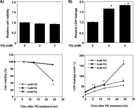
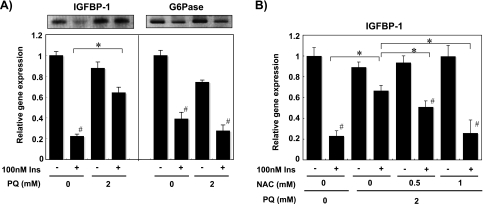
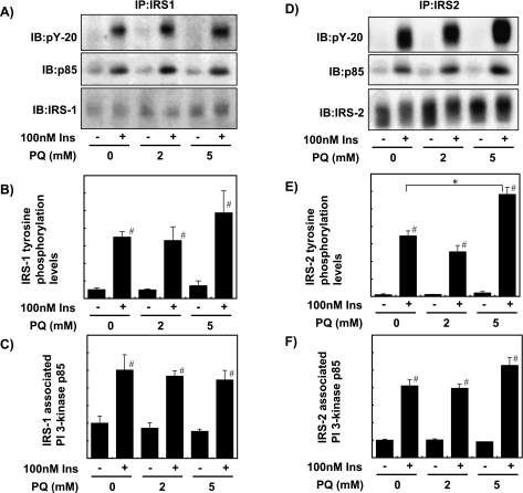
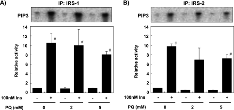
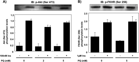
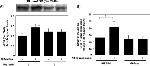
Oxidative stress is thought to play a role in the development of insulin resistance. In order to elucidate the molecular effect of oxidative stress on liver insulin signaling, we analyzed the effect of paraquat (1,1-dimethyl-4,4-dipyridynium; PQ)-derived oxidative stress on the expression of insulin-dependent genes and activation of liver insulin signaling pathway. Incubation of primary cultured rat hepatocytes with 2 mM PQ for 6 h impaired the suppressive effect of insulin on insulin-like growth factor-binding protein-1 (IGFBP-1) gene expression, but did not influence glucose-6-phosphatase gene expression. Insulin-dependent phosphorylation or activation of insulin receptor, insulin receptor substrate-1 and -2, phosphatidylinositol 3-kinase, Akt and forkhead in rhabdomyosarcoma were not affected by PQ pre-treatment. In contrast, PQ treatment impaired insulin-dependent phosphorylation of mammalian target of rapamycin (mTOR). These results indicate that PQ-induced oxidative stress impairs insulin-dependent mTOR activation and that this impairment probably causes inhibition of insulin-dependent repression of IGFBP-1 expression.
Keywords: IGFBP-1; hepatocyte; insulin; mTOR; paraquat.
Figures
Similar articles
-
Insulin regulation of hepatic insulin-like growth factor-binding protein-1 (IGFBP-1) gene expression and mammalian target of rapamycin (mTOR) signalling is impaired by the presence of hydrogen peroxide.Biochem J. 2002 Jul 15;365(Pt 2):537-45. doi: 10.1042/BJ20020266. Biochem J. 2002. PMID: 11942857 Free PMC article.
-
Ligustrazin increases lung cell autophagy and ameliorates paraquat-induced pulmonary fibrosis by inhibiting PI3K/Akt/mTOR and hedgehog signalling via increasing miR-193a expression.BMC Pulm Med. 2019 Feb 11;19(1):35. doi: 10.1186/s12890-019-0799-5. BMC Pulm Med. 2019. PMID: 30744607 Free PMC article.
-
Effect of various radical generators on insulin-dependent regulation of hepatic gene expression.Biosci Biotechnol Biochem. 2007 Jan;71(1):16-22. doi: 10.1271/bbb.60016. Epub 2007 Jan 7. Biosci Biotechnol Biochem. 2007. PMID: 17213664
-
Paraquat induces apoptosis in human lymphocytes: protective and rescue effects of glucose, cannabinoids and insulin-like growth factor-1.Growth Factors. 2008 Feb;26(1):49-60. doi: 10.1080/08977190801984205. Growth Factors. 2008. PMID: 18365879
-
New insights into Notch1 regulation of the PI3K-AKT-mTOR1 signaling axis: targeted therapy of γ-secretase inhibitor resistant T-cell acute lymphoblastic leukemia.Cell Signal. 2014 Jan;26(1):149-61. doi: 10.1016/j.cellsig.2013.09.021. Epub 2013 Oct 16. Cell Signal. 2014. PMID: 24140475 Review.
Cited by
-
Protective Effect of Fenofibrate on Oxidative Stress-Induced Apoptosis in Retinal-Choroidal Vascular Endothelial Cells: Implication for Diabetic Retinopathy Treatment.Antioxidants (Basel). 2020 Aug 5;9(8):712. doi: 10.3390/antiox9080712. Antioxidants (Basel). 2020. PMID: 32764528 Free PMC article.
-
Insulin Signaling Disruption and INF-γ Upregulation Induce Aβ1-42 and Hyperphosphorylated-Tau Proteins Synthesis and Cell Death after Paraquat Treatment of Primary Hippocampal Cells.Chem Res Toxicol. 2022 Dec 19;35(12):2214-2218. doi: 10.1021/acs.chemrestox.2c00278. Epub 2022 Nov 17. Chem Res Toxicol. 2022. PMID: 36394833 Free PMC article.
-
Herbicide-related health risks: key mechanisms and a guide to mitigation strategies.J Occup Med Toxicol. 2025 Feb 25;20(1):6. doi: 10.1186/s12995-025-00448-7. J Occup Med Toxicol. 2025. PMID: 40001182 Free PMC article. Review.
-
Nrf2 deficiency improves glucose tolerance in mice fed a high-fat diet.Toxicol Appl Pharmacol. 2012 Nov 1;264(3):305-14. doi: 10.1016/j.taap.2012.09.014. Epub 2012 Sep 24. Toxicol Appl Pharmacol. 2012. PMID: 23017736 Free PMC article.
References
-
- Dandona P., Thusu K., Cook S., Snyder B., Makowski J., Armstrong D., Nicotera T. Oxidative damage to DNA in diabetes mellitus. Lancet. 1996;347:444–445. - PubMed
-
- Nourooz Z.J., Rahimi A., Tajaddini S.J., Tritschler H., Rosen P., Halliwell B., Betteridge D.J. Relationship between plasma measures of oxidative stress and metabolic control in NIDDM. Diabetologia. 1997;34:171–175. - PubMed
-
- Baynes J.W., Thorpe S.R. Role of oxidative stress in diabetic complications; a new perspective on an old paradigm. Diabetes. 1999;48:1–9. - PubMed
-
- Paolisso G., D’Amore A., Giugliano D., Ceriello A., Varrricchio M., D’Onofrio F. Pharmacologic doses of vitamin E improve insulin action in healthy subjects and non-insulin-dependent diabetic patients. Am. J. Clin. Nutr. 1993;57:650–656. - PubMed
-
- Faure P., Rossini E., Lafond J.L., Richard M.J., Favier A., Halimi S. Vitamin E improves the free radical defence system potential and insulin sensitivity of rats fed high fructose diets. J. Nutr. 1997;127:103–107. - PubMed
LinkOut - more resources
Full Text Sources
Molecular Biology Databases
Research Materials
Miscellaneous