. 2010 Jul;59(7):1059-71.
doi: 10.1007/s00262-010-0831-3. Epub 2010 Mar 9.

Antitumor activity of type I and type III interferons in BNL hepatoma model

Affiliations

Antitumor activity of type I and type III interferons in BNL hepatoma model

Walid Abushahba et al. Cancer Immunol Immunother. 2010 Jul.

Abstract

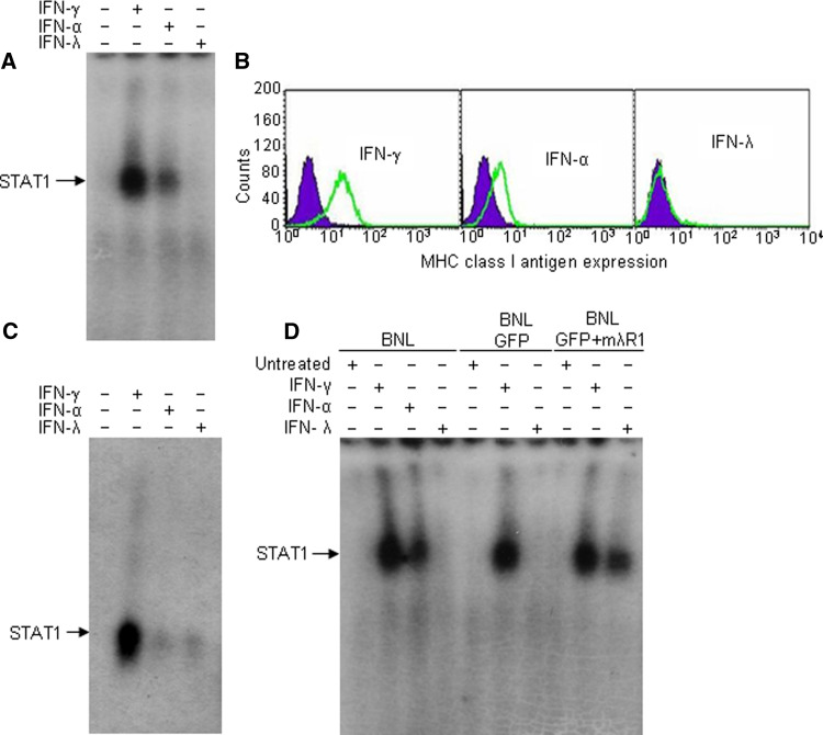
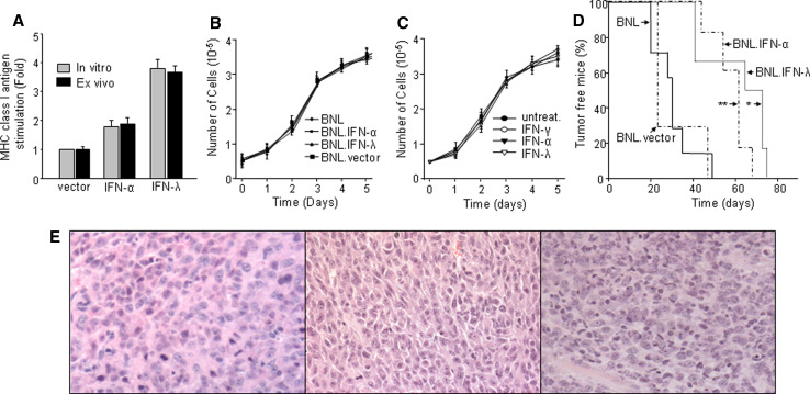
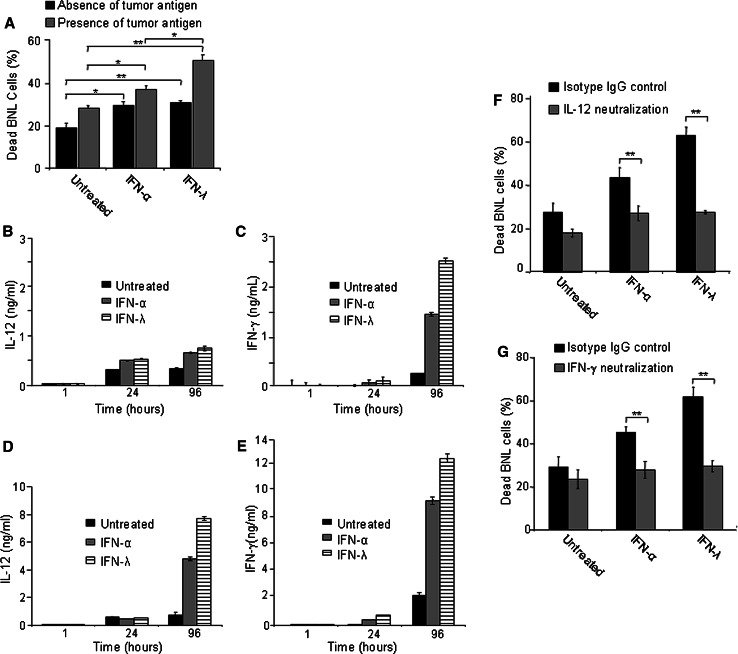
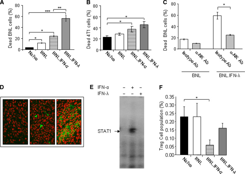
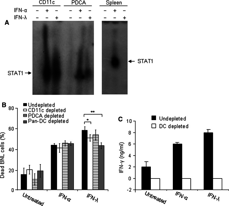
Hepatocellular carcinoma (HCC) occurs most commonly secondary to cirrhosis due to chronic hepatitis C or B virus (HCV/HBV) infections. Type I interferon (IFN-alpha) treatment of chronic HCV/HBV infections reduces the incidence of HCC in cirrhotic patients. However, IFN-alpha toxicity limits its tolerability and efficacy highlighting a need for better therapeutic treatments. A recently discovered type III IFN (IFN-lambda) has been shown to possess antiviral properties against HCV and HBV in vitro. In phase I clinical trials, IFN-lambda treatment did not cause significant adverse reactions. Using a gene therapy approach, we compared the antitumor properties of IFN-alpha and IFN-lambda in a transplantable hepatoma model of HCC. BALB/c mice were inoculated with syngeneic BNL hepatoma cells, or BNL cells expressing IFN-lambda (BNL.IFN-lambda cells) or IFN-alpha (BNL.IFN-alpha cells). Despite the lack of antiproliferative activity of IFNs on BNL cells, both BNL.IFN-lambda and BNL.IFN-alpha cells displayed retarded growth kinetics in vivo. Depletion of NK cells from splenocytes inhibited splenocyte-mediated cytotoxicity, demonstrating that NK cells play a role in IFN-induced antitumor responses. However, isolated NK cells did not respond directly to IFN-lambda. There was also a marked NK cell infiltration in IFN-lambda producing tumors. In addition, IFN-lambda and, to a lesser extent, IFN-alpha enhanced immunocytotoxicity of splenocytes primed with irradiated BNL cells. Splenocyte cytotoxicity against BNL cells was dependent on IL-12 and IFN-gamma, and mediated by dendritic cells. In contrast to NK cells, isolated from spleen CD11c+ and mPDCA+ dendritic cells responded directly to IFN-lambda. The antitumor activities of IFN-lambda against hepatoma, in combination with HCV and HBV antiviral activities warrant further investigation into the clinical use of IFN-lambda to prevent HCC in HCV/HBV-infected cirrhotic patients, as well as to treat liver cancer.


Figures

Fig. 1
Fig. 1
IFN response in BNL cells. STAT activation in murine hepatoma BNL cells (a) and in primary mouse hepatocytes (c) in response to mouse IFN-γ, IFN-α or IFN-λ was evaluated by EMSA with a GAS probe. Cells were treated for 15 min with IFNs (10 ng/ml), lysed and STAT activation was detected by EMSA. Position of STAT DNA-binding-complexes in EMSA is indicated by the arrow. The results are representative of three independent experiments. b The level of MHC class I antigen expression was determined by flow cytometry in BNL cells untreated (black filled histograms) or treated for 72 h with recombinant mIFN-α, mIFN-λ2 or mIFN-γ (10 ng/ml, gray open histograms). The data shown are representative of three independent experiments. d BNL cells were transiently transfected with mouse IFN-λR1, treated with IFNs and STAT1 activation was determined by EMSA
Fig. 2
Fig. 2
Constitutive expression of IFNs in BNL cells and its effects on tumor growth. a B16 melanoma cells were treated for 72 h with either conditioned medium from BNL.vector, BNL.IFN-λ or BNL.IFN-α cells (the pre-implantation cell cultures; gray bars), or with conditioned medium from BNL.vector, BNL.IFN-λ or BNL.IFN-α ex vivo cell cultures [BNL.vector, BNL.IFN-λ or BNL.IFN-α tumors were extracted from mice (n = 5), tumor cells were isolated and grown ex vivo; black bars]. The level of MHC class I antigen expression was determined by flow cytometry and the fold increase over the basal level of MHC class I antigen expression in untreated B16 cells was calculated. Data shown are representative of two independent experiments and presented as the mean ± SD (n = 5 per data point). b Growth rate of parental BNL, BNL.vector, BNL.IFN-α, or BNL.IFN-λ cells was assessed by growing an equal number of cells (5 × 104) in each well and counting the cells at days 1, 2, 3, 4, and 5. These experiments were performed in triplicate. Data are presented as the mean ± SD. c Antiproliferative activity of recombinant murine IFNs was evaluated on BNL cells. An equal number of BNL cells (5 × 104) was plated in all wells, the cells were left untreated or treated with either mIFN-γ, mIFN-α or mIFN-λ2 (10 ng/ml). Every day for 5 days, cells were collected and counted. These experiments were performed in triplicate. Data are presented as the mean ± SD. d Tumorigenicity of parental and modified IFN-producing BNL cells was evaluated in immunocompetent syngeneic BALB/c mice. Mice (n = 8) were injected s.c. in the flank with 106 BNL, BNL.vector, BNL.IFN-λ or BNL.IFN-α cells, and tumor development was monitored every other day by palpation of the injection site. The results are representative of two independent experiments. Data are shown as percentage of tumor free mice. BNL.vector versus BNL.IFN-λ *P < 0.05; BNL.vector versus BNL.IFN-α **P = 0.09. e H&E staining followed by light microscopy (×10) was performed on BNL.parental (left), BNL.IFN-α (middle) or BNL.IFN-λ tumors (right)
Fig. 3
Fig. 3
IFN-induced immunocytotoxicity and production of IL-12 and IFN-γ. a Splenocytes (106) were isolated from naive mice (n = 3) and treated with IFNs in the presence or absence of irradiated (105) BNL cells (4 day culture). On day 4, splenocytes were collected and co-cultured with fresh (105) target BNL cells for 24 h. The cells were harvested, stained with propidium iodide, and the amount of dead target BNL tumor cells was assessed by FACS. Dead parental BNL cell numbers (%) are shown as the mean ± SD (n = 3 per data point). Results are representative of two independent experiments. *P < 0.05; **P < 0.01. b–e Conditioned media of splenocytes treated with IFNs in the absence (b, c) or presence (d, e) of tumor antigens were collected at 1, 24, and 96 h of the treatment and the levels of IL-12 (b, d) and IFN-γ (c, e) in the conditioned media were analyzed. Data are presented as the mean value of triplicate wells ± SD. f, g Untreated or IFN-treated splenocyte cultures were incubated in the presence of irradiated BNL cells with 20 μg/mL of purified anti-IL-12p40 (f), purified anti-mouse IFN-γ (g) or isotype-matched rat IgG and splenocyte immunocytotoxicity was measured after the 4 day treatment as described in a. Dead parental BNL cell numbers (%) are shown as the mean ± SD (n = 3 per data point). All results are representative of two independent experiments. F and G: **P < 0.01
Fig. 4
Fig. 4
IFN-λ triggers a substantial immunocytotoxic response against target tumor cells and the involvement of NK cells. a, b Splenocytes were isolated from naive mice or mice (n = 5) injected with either parental BNL, BNL.IFN-α, or BNL.IFN-λ cells, and their cytotoxicity was tested against parental BNL tumor cells (a) or heterologous 4T1 breast carcinoma cells (b). Dead parental BNL (a) or 4T1 (b) cell numbers (%) are shown as the mean ± SD [n = 5 (a) and n = 3 (b) per data point]. Results in (a) are representative of two independent experiments. *P < 0.05, **P < 0.01, ***P < 0.001. c NK cells were depleted from splenocytes isolated from mice injected with parental BNL or BNL.IFN-λ cells with the use of a biotinylated rat monoclonal Ab against NK or with isotype-matched rat IgM as a control. Cytotoxicity of splenocytes against parental BNL cells was evaluated in vitro as described above. Dead parental BNL cell numbers (%) are shown as the mean ± SD (n = 3 per data point). Results are representative of three independent experiments. *P < 0.05. d Immunohistochemical staining of CD49b+ NK cells (green fluorescence) was performed in BNL.parental (left), BNL.IFN-α (middle) or BNL.IFN-λ tumors (right). Samples were examined in a confocal fluorescent microscope at ×250. e NK cells were purified from spleen of mice which received BNL.IFN-λ cells with the use of magnetic beads as described in “Materials and methods”. NK cells were treated with IFN-α or IFN-λ (10 ng/ml) for 15 min, lysed and STAT1 activation was detected in cellular extracts by EMSA. The EMSA is representative of two independent experiments. f PBMCs were isolated from naive mice or mice injected with either parental BNL cells, BNL.IFN-α, or BNL.IFN-λ cells, and the number of circulating CD4+ CD25+ Foxp3+ Tregs in the lymphoid gate population (%) was determined by flow cytometry and shown as the mean ± SD (n = 3 per data point). *P < 0.05
Fig. 5
Fig. 5
Involvement of DCs in IFN-λ-induced immunocytotoxic response. a CD11c+ and mPDCA+ DCs were purified from mouse spleens using magnetic MACS microbeads (as described in the “Materials and methods”). DCs and total splenocytes were treated with IFN-α or IFN-λ (10 ng/ml) for 15 min, lysed and STAT1 activation was detected in cellular extracts by EMSA. b In vitro depletion of CD11c+, mPDCA+, or both populations in splenocytes (106) was performed (as described in the “Materials and methods”). Splenocytes were harvested from naive mice (n = 3) and treated with IFNs in the presence or absence of irradiated (105) BNL cells (4 day culture). On day 4, splenocytes were collected and co-cultured with fresh (105) target BNL cells for 24 h. The cells were harvested, stained with propidium iodide, and the amount of dead target BNL tumor cells was assessed by FACS. Dead parental BNL cell numbers (%) are shown as the mean ± SD (n = 3 per data point). *P < 0.05, **P < 0.01. c Conditioned media of splenocytes (n = 3) treated with IFNs in (a) were collected after 4 days of treatment and the levels of IFN-γ in the conditioned media were analyzed. Data are presented as the mean value of triplicate wells ± SD

References

    1. Sherman M. Hepatocellular carcinoma: epidemiology, risk factors, and screening. Semin Liver Dis. 2005;25:143–154. doi: 10.1055/s-2005-871194. - DOI - PubMed
    1. Farazi PA, DePinho RA. Hepatocellular carcinoma pathogenesis: from genes to environment. Nat Rev Cancer. 2006;6:674–687. doi: 10.1038/nrc1934. - DOI - PubMed
    1. Lau WY, Lai EC. Hepatocellular carcinoma: current management and recent advances. Hepatobiliary Pancreat Dis Int. 2008;7:237–257. - PubMed
    1. Taketomi A, Soejima Y, Yoshizumi T, Uchiyama H, Yamashita Y, Maehara Y. Liver transplantation for hepatocellular carcinoma. J Hepatobiliary Pancreat Surg. 2008;15:124–130. doi: 10.1007/s00534-007-1296-4. - DOI - PubMed
    1. Mazzaferro V, Chun YS, Poon RT, Schwartz ME, Yao FY, Marsh JW, Bhoori S, Lee SG. Liver transplantation for hepatocellular carcinoma. Ann Surg Oncol. 2008;15:1001–1007. doi: 10.1245/s10434-007-9559-5. - DOI - PMC - PubMed

Publication types

MeSH terms