

Combined detection of Her2/neu gene amplification and protein overexpression in effusions from patients with breast and ovarian cancer
- PMID: 20217132
- PMCID: PMC11827919
- DOI: 10.1007/s00432-010-0790-2
Combined detection of Her2/neu gene amplification and protein overexpression in effusions from patients with breast and ovarian cancer
Abstract
Purpose: Her2/neu protein overexpression and gene amplification is found in 20-30% of breast cancer patients and correlates with poor clinical outcome. Patients who profit from anti-Her2/neu- therapy are routinely selected by examination of tumour specimens using immunohistochemistry (IHC) and fluorescence in situ hybridisation (FISH) separately. Many studies found a good correlation between both methods for score 1+ samples and for score 3+ samples, but not for score 2+ samples. In this study, we examined pleural and ascitic effusions with a combined approach using IHC and FISH on the same cells, under the following aspects: (1) frequency of Her2/neu protein expression and gene amplification in effusions; (2) correlation between score of protein expression and gene amplification; (3) impact of chromosome 17 polyploidy on Her2/neu protein expression.
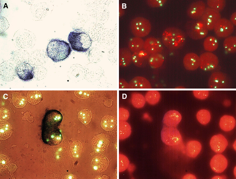
Methods: We examined 31 effusions from patients with breast cancer and 4 effusions from patients with ovarian cancer. Cytospins were analysed by IHC using two anti-Her2/neu antibodies and subsequently analysed by FISH with Her2/neu/CEP17 probes. Amplification was defined as: (1) Her2/neu gene copy number of more than 4 and (2) Her2/neu/CEP17 ratio of 2.0 or greater.
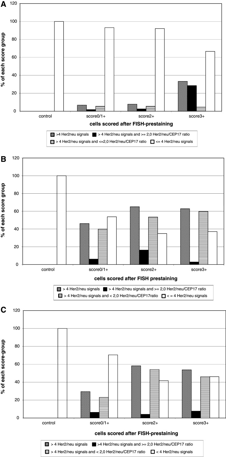
Results: A system combining IHC and FISH was developed and 35 effusion specimens were examined. As much as 25 of them were scored as positive. All of them contained cells with heterogeneous scores. A total of 18 of the samples contained cells with scores that ranged from 0 to 3+. In the other samples scores ranged from 0 to 1+ or 0 to 2+. Cells were analysed for Her2/neu gene amplification and chromosome 17 ploidy with regard to their scores. As much as 15 of all samples had mean Her2/neu copy numbers of >4, but only 12% (n = 3) of the positive samples were amplified according to Her2/neu/CEP17 ratio. Only 22, 9% (n = 8) were polyploid (mean CEP17 > 4); but 65, 7% (n = 23) of all specimens contained single polyploid cells. In some cases up to 100% of the score 3+ cells showed chromosome 17 polyploidy. Here protein overexpression might be caused by polyploidy rather than by gene amplification. In some samples, we found single cells with gene amplification but without protein expression and cells without amplification but with protein overexpression.
Conclusion: The combination of IHC and FISH allows a differentiated analysis of single cells, which is especially important for effusions that are composed of heterogeneous cells. Therefore, cells with high gene amplification and/or protein overexpression can be detected and analysed even if their amount in the sample is small. Chromosome 17 polyploidy is important in some cases but this should be further examined on a larger series.
Figures

Similar articles
-
Topoisomerase IIalpha expression rather than gene amplification predicts responsiveness of adjuvant anthracycline-based chemotherapy in women with primary breast cancer.J Cancer Res Clin Oncol. 2010 Jul;136(7):1029-37. doi: 10.1007/s00432-009-0748-4. Epub 2010 Jan 6. J Cancer Res Clin Oncol. 2010. PMID: 20052594 Free PMC article.
-
Home treatment for mental health problems: a systematic review.Health Technol Assess. 2001;5(15):1-139. doi: 10.3310/hta5150. Health Technol Assess. 2001. PMID: 11532236
-
A rapid and systematic review of the clinical effectiveness and cost-effectiveness of paclitaxel, docetaxel, gemcitabine and vinorelbine in non-small-cell lung cancer.Health Technol Assess. 2001;5(32):1-195. doi: 10.3310/hta5320. Health Technol Assess. 2001. PMID: 12065068
-
Cost-effectiveness of using prognostic information to select women with breast cancer for adjuvant systemic therapy.Health Technol Assess. 2006 Sep;10(34):iii-iv, ix-xi, 1-204. doi: 10.3310/hta10340. Health Technol Assess. 2006. PMID: 16959170
-
Comparison of Two Modern Survival Prediction Tools, SORG-MLA and METSSS, in Patients With Symptomatic Long-bone Metastases Who Underwent Local Treatment With Surgery Followed by Radiotherapy and With Radiotherapy Alone.Clin Orthop Relat Res. 2024 Dec 1;482(12):2193-2208. doi: 10.1097/CORR.0000000000003185. Epub 2024 Jul 23. Clin Orthop Relat Res. 2024. PMID: 39051924
Cited by
-
Relationship between HER2 and JAK/STAT-SOCS3 signaling pathway and clinicopathological features and prognosis of ovarian cancer.Cancer Biol Ther. 2017 May 4;18(5):314-322. doi: 10.1080/15384047.2017.1310343. Epub 2017 Apr 27. Cancer Biol Ther. 2017. PMID: 28448787 Free PMC article.
-
Advances in the Application of Radionuclide-Labeled HER2 Affibody for the Diagnosis and Treatment of Ovarian Cancer.Front Oncol. 2022 Jun 15;12:917439. doi: 10.3389/fonc.2022.917439. eCollection 2022. Front Oncol. 2022. PMID: 35785201 Free PMC article. Review.
-
A Novel, Personalized Drug-Screening System for Platinum-Resistant Ovarian Cancer Patients: A Preliminary Clinical Report.Cancer Manag Res. 2021 Mar 29;13:2849-2867. doi: 10.2147/CMAR.S276799. eCollection 2021. Cancer Manag Res. 2021. PMID: 33833569 Free PMC article.
-
Pre-analytical issues in effusion cytology.Pleura Peritoneum. 2016 Mar 1;1(1):45-56. doi: 10.1515/pp-2016-0001. Epub 2016 Apr 12. Pleura Peritoneum. 2016. PMID: 30911607 Free PMC article. Review.
-
Targeted Nanocarrier-Based Drug Delivery Strategies for Improving the Therapeutic Efficacy of PARP Inhibitors against Ovarian Cancer.Int J Mol Sci. 2024 Jul 30;25(15):8304. doi: 10.3390/ijms25158304. Int J Mol Sci. 2024. PMID: 39125873 Free PMC article. Review.
References
-
- Acs G, Wang L, Raghunath PN, Salscheider MA, Zhang PJ (2003) Role of different immunostaining patterns in Herceptest interpretation and criteria for gene amplification as determined by fluorescence in situ hybridization. Appl Immunohistochem Mol Morphol 11(3):222–229 - PubMed
-
- Bermont L, Algros MP, Baron MH, Adessi GL (2000) Relevance of p185 Her-2/neu oncoprotein quantification in human primary breast carcinoma. Breast Cancer Res Treat 63:163–169 - PubMed
-
- Bilous M, Ades C, Armes J et al (2003) Predicting the HER2 status of breast cancer from basic histopathology data, an analysis of 1500 breast cancers as part of the HER 2000 international study. Breast 12:92–98 - PubMed
-
- Bofin AM et al (2003) TOP2A and HER-2 gene amplification in fine needle aspirates from breast carcinomas. Cytopathology 14:314–319 - PubMed
MeSH terms
Substances
LinkOut - more resources
Full Text Sources
Other Literature Sources
Medical
Research Materials
Miscellaneous
