Review
. 2010 Aug;67(15):2533-50.
doi: 10.1007/s00018-010-0318-6. Epub 2010 Mar 9.

The neonatal Fc receptor as therapeutic target in IgG-mediated autoimmune diseases

Affiliations
Review

The neonatal Fc receptor as therapeutic target in IgG-mediated autoimmune diseases

Alina Sesarman et al. Cell Mol Life Sci. 2010 Aug.

Abstract

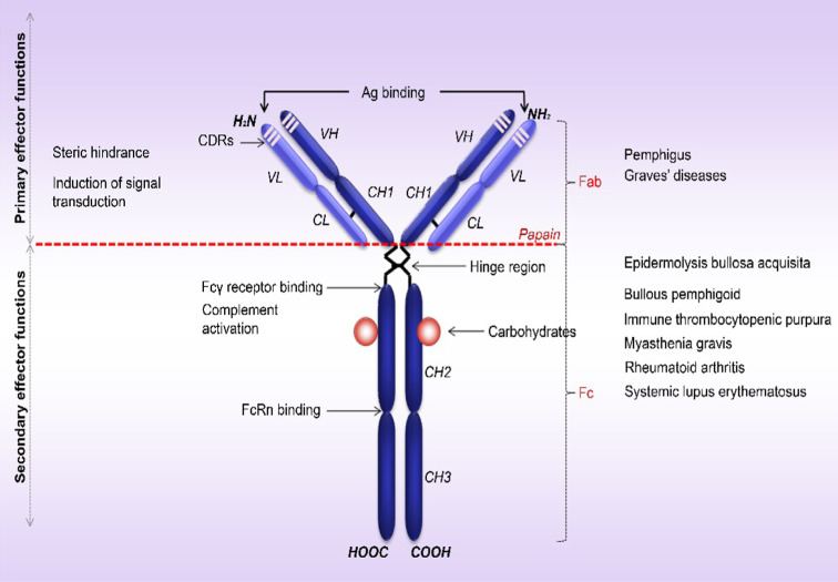
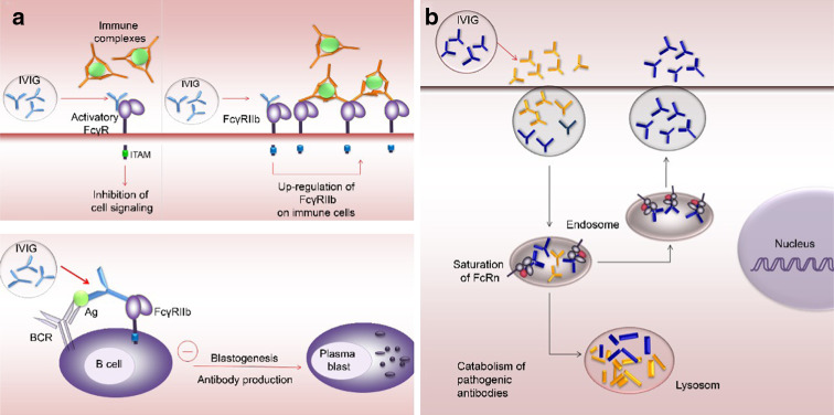
Therapy approaches based on lowering levels of pathogenic autoantibodies represent rational, effective, and safe treatment modalities of autoimmune diseases. The neonatal Fc receptor (FcRn) is a major factor regulating the serum levels of IgG antibodies. While FcRn-mediated half-life extension is beneficial for IgG antibody responses against pathogens, it also prolongs the serum half-life of IgG autoantibodies and thus promotes tissue damage in autoimmune diseases. In the present review article, we examine current evidence on the relevance of FcRn in maintaining high autoantibody levels and discuss FcRn-targeted therapeutic approaches. Further investigation of the FcRn-IgG interaction will not only provide mechanistic insights into the receptor function, but should also greatly facilitate the design of therapeutics combining optimal pharmacokinetic properties with the appropriate antibody effector functions in autoimmune diseases.


Figures

Fig. 1
Fig. 1
Structure of human IgG and mechanisms of autoantibody-induced tissue damage. Human IgG molecule contains two identical (H) heavy chains (50 kDa) covalently linked to two (L) light chains (23 kDa) by disulfide bonds. Each chain is organized in constant (C) and variable (V) structural domains. The heavy chain has three constant domains (CH1, CH2, CH3) and one variable domain (VH), while the associated light chain has one constant (CL) and one variable domain (VL). The variable domains of the heavy chain (VH) and the light chain (VL) have three complementarity determining regions (CDRs; CDR1, CDR2 and CDR3) with major contribution to the antigen binding site. Papain cleaves the IgG molecule above the hinge region, giving rise to two Fab fragments (fragment, antigen-binding) involved in antigen recognition and one Fc fragment (fragment, crystallizable), which binds to the C1q, FcγRs and FcRn and mediates the activation of complement and/or immune cells and IgG trafficking at various sites, respectively
Fig. 2
Fig. 2
Human Fc receptors for IgG. The Fc region of IgG binds several types of Fc receptors, including activatory and inhibitory Fcγ receptors and the neonatal Fc receptor (FcRn). a Human Fcγ receptors, termed FcγRI (CD64), FcγRII (CD32), FcγRIII (CD16) are cell-surface molecules primarily expressed on immune cells. Structurally, these receptors share a highly homologous extracellular region composed of immunoglobulin-like domains, but they show major differences in the organization of the transmembrane and cytoplasmic regions, hence their different effector functions. With the exception of FcγRIIb, which is associated with the cytoplasmic immune receptor thyrosine-based inhibtion motif (ITIM), FcγRI, and FcγRIIIa associate with the Fc-Receptor γ-chain (FcRγ-chain) that possess a cytoplasmic immune receptor thyrosine-based activation motif (ITAM), whereas FcγRIIa and FcγRIIc contain their own cytoplasmic ITAM. The ITAM- and the ITIM-containing FcγR elicit different and at times opposing types of intracellular signaling mechanisms. b The neonatal Fc receptor for IgG, is structurally similar to the MHC-class I molecules, rather then the Fcγ family, consisting of a transmembrane glycosylated alpha chain non-covalently coupled to β2-microglobulin. Unlike classical Fcγ receptors, FcRn binds IgG at low pH, mediating the transfer of these antibodies from mother to young, and preserving it in the circulation during the adult life. FcRn is expressed in endothelial, epithelial, and hematopoietic cells. In myeloid cells, in addition to contributing to IgG homeostasis, FcRn modulates IgG-mediated phagocytosis and antigen presentation
Fig. 3
Fig. 3
FcRn functions. a In adult life, FcRn is responsible for the regulation of IgG persistence in the circulation. IgGs are internalized by endothelial cells by fluid phase pinocytosis. At the low pH of the acidic endosomes FcRn binds IgG, transports it back to the cell surface where, due to the neutral pH, complex stability decreases and IgG is released/recycled. Unbound IgG is catabolized in the lysosomes. FcRn also controls the transfer of antibodies from b mother’s milk to neonates in the gut and from c maternal plasma through the placenta to fetus in most mammals. IgG-FcRn interaction is pH dependent, occurring efficiently at an acidic pH 6 (as found in intestinal lumen or endosomes). Binding of FcRn to IgG becomes progressively weaker as the pH raises to near neutral, until at around pH 7.4 it is negligible. Receptor-bound IgGs are then transcytosed across the epithelial intestinal cells (b) or syncitiotrophoblasts (c) being released from this complex in the neonatal or fetus tissues/bloodstream upon exposure to the neutral pH-7.4 of these millieu
Fig. 4
Fig. 4
FcRn-deficiency protects from tissue injury in experimental epidermolysis bullosa acquisita. Mice were injected every second day with 5 mg of pathogenic rabbit antibodies against murine type VII collagen (mCVII) [72]. Characteristic blisters, erosions, and crusts were detected 18 days after the first injection in a wild-type animals, while no cutaneous lesions were observed in b FcRn-/- mice. Histological and immunopathological examination of skin biopsies obtained from wild-type mice showed c subepidermal blisters and accumulation of neutrophils as wells as deposition of e IgG and g C3 at the dermal-epidermal junction. d No histological changes were detected in an FcRn-/- mouse injected with rabbit anti-collagen VII IgG. Immunofluorescence analysis of skin biopsies of the FcRn-/- mice revealed positive staining for f rabbit IgG and discrete focal deposition of h murine C3 at the dermal–epidermal junction (magnification ×200). i Serum samples obtained from experimental animals were analyzed by ELISA to measure the levels of antibodies to type VII collagen. Compared to wild-type mice, FcRn-/- mice showed significantly lower levels of circulating pathogenic IgG
Fig. 5
Fig. 5
Major Fc-dependent mechanisms of action for IVIG in IgG-mediated autoimmune diseases. a Blocking of activating FcγRs and up-regulation of inhibitory FcγRs. In IgG-mediated inflammatory diseases, activation of FcγR expressed on immune cells may essentially contribute to tissue destruction. Antibodies found in IVIG compete with antigen-IgG complexes for binding the activating FcγRs, thus limiting or inhibiting their ability to trigger cell signaling events. In addition, IVIG interaction with the FcγRIIb increases its expression on immune effector cells, therefore, triggering the activating FcγR requires higher concentration of immune complexes. This “inhibition” appears to be mainly responsible for the anti-inflammatory activity proposed of IVIG. Upon co-ligation of the B-cell antigen receptor (BCR) with the FcγRIIb, IVIG activates an inhibitory signaling pathway on B lymphocytes, impairing blastogenesis and IgG synthesis. b Saturation of FcRn. IgG in IVIG formulations binds to and saturates the FcRn leading to accelerated the catabolism of pathogenic antibodies and thereby lowering their levels in the circulation below the threshold required for disease progression

References

    1. Sinha AA, Lopez MT, McDevitt HO. Autoimmune diseases: the failure of self tolerance. Science. 1990;248:1380–1388. - PubMed
    1. Bach J. The effect of infections on susceptibility to autoimmune and allergic diseases. N Engl J Med. 2002;347:911–920. - PubMed
    1. Drachman DB. How to recognize an antibody-mediated autoimmune disease: criteria. Res Publ Assoc Res Nerv Ment Dis. 1990;68:183–186. - PubMed
    1. Rose NR, Bona C. Defining criteria for autoimmune diseases (Witebsky’s postulates revisited) Immunol Today. 1993;14:426–430. - PubMed
    1. Stassi G, De Maria R. Autoimmune thyroid disease: new models of cell death in autoimmunity. Nat Rev Immunol. 2002;2:195–204. - PubMed

Publication types