. 2010 Aug;59(8):659-66.
doi: 10.1007/s00011-010-0178-4. Epub 2010 Mar 9.

C5a promotes migration, proliferation, and vessel formation in endothelial cells

Affiliations

C5a promotes migration, proliferation, and vessel formation in endothelial cells

Ryuji Kurihara et al. Inflamm Res. 2010 Aug.

Abstract

Objectives: The goal of this paper is to investigate the effects of activated complement C5a on vascular endothelium during vessel formation.

Methods: A human microvascular endothelial cell line (HMEC-1) derived from post-capillary venules in skin was used to measure DNA synthesis, proliferation and cell-cycle progression. In vitro ring-shaped formation by the cells was assessed by using type I collagen gel matrix and a cell-migration assay using the Chemotaxicell chamber. A Matrigel plug assay was performed to confirm the effect of C5a in vivo.

Results: C5a progressed the cell cycle of HMEC-1 into G2/M phases, and induced DNA synthesis and proliferation in a dose-dependent manner. C5a efficiently induced migration and ring-shaped structure formation both in vitro and in vivo. Furthermore, a C5a receptor antagonist (W-54011) suppressed all HMEC-1 activities including proliferation and migration.

Conclusions: Proliferation, migration, and ring-shaped formation by HMEC-1 cells was induced by C5a. The actions were efficiently inhibited by a specific antagonist against C5a. Our results implicated C5a in vessel formation and as a potent target for management of inflammatory diseases.


Figures
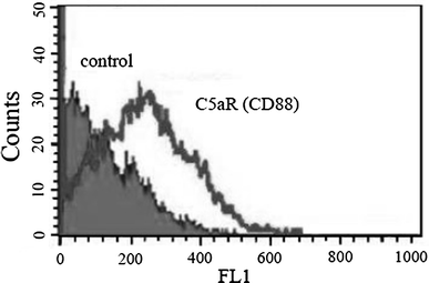
Fig. 1
Fig. 1
Expression of C5aR (CD88) on human microvascular endothelial cells (HMEC-1). Flow cytometry results showing C5aR expression (outlined area) on HMEC-1 cells without stimulation. Dark areas indicate control
Fig. 2
Fig. 2
Cell proliferation induced by C5a. [3H]-thymidine incorporation was measured with a liquid scintillation counter 48 h after adding the various concentrations of C5a, C5, or C3a to the HMEC-1 suspension. a C5a increased HMEC-1 proliferation in a dose-dependent manner, whereas C5 (b) and C3a (c) did not induce proliferation. d HMEC-1 was stimulated with either 10 nM C5a in the presence of various concentrations of C5a receptor antagonist W-54011 (closed column) or W-54011 alone (open column). The addition of W-54011 inhibited cell proliferation induced by C5a 10 nM in a dose-dependent manner, whereas W-54011 alone had no effect. Bars represent mean ± SEM (n = 5). **P < 0.01 by Dunnett’s multiple comparison test
Fig. 3
Fig. 3
Cell cycle progression induced by C5a. HMEC-1 cultures were incubated with 10 nM C5a or the indicated concentrations of C5 and C3a. Cell cycle phases were determined by flow cytometry with PI staining enabling the determination of cells in the G0/G1 and S/G2/M phases. a Cells in the S/G2/M phases advanced significantly when stimulated with 10 nM C5a for 48 h, and this effect was suppressed by the addition of 10 nM W-54011. b C5a promoted cell cycle progression in a dose-dependent manner, C5 (c) and C3a (d) had no effect on the stage of cell cycle. e The indicated concentrations of W-54011 were added in the presence of 10 nM C5a (closed bars) or without C5a (open bars). W-54011 suppressed the effect of C5a on cell cycle whereas W-54011 alone had no effect. Bars represent mean ± SEM (n = 5). *P < 0.05 and **P < 0.01 by Dunnett’s multiple comparison test
Fig. 4
Fig. 4
Cell viability was measured using the Tetra Color One assay. The optical density of each well was measured at 450 nm. Phosphate-buffered saline (PBS) alone was added to control samples. a C5a increased the number of viable HMEC-1 cells at 1 and 10 nM, while C5 (b) and C3a (c) had no such effects. d W-54011 inhibited the C5a-augmented viable cell count (closed bars), but had no effect when added alone (open bars). Bars represent mean ± SEM (n = 5). **P < 0.01 by Dunnett’s multiple comparison test
Fig. 5
Fig. 5
Formation of ring-shaped structures in vitro. a Cells incubated in the presence of phosphate-buffered saline (PBS) alone showed minimal ring-shaped arrangements of the endothelial cells. Fibroblast growth factor 2 (FGF-2; 1 ng/mL) induced slight ring-structure formation, while 10 nM C5a dramatically increased the number of ring-shape structures. This C5a-induced effect was inhibited by W-54011. Original magnification: ×40. b C5a increased the number of ring-shaped structures formed by HMEC-1 in a dose-dependent manner compared with FGF-2 alone. C5 and C3a did not induce the formation of ring-shaped structures. c W-54011 inhibited ring-shaped structure formation in a dose-dependent manner (closed bars) whereas W-54011 alone had no effect (open bars). Bars represent mean ± SEM (n = 5). **P < 0.01 by Dunnett’s multiple comparison test
Fig. 6
Fig. 6
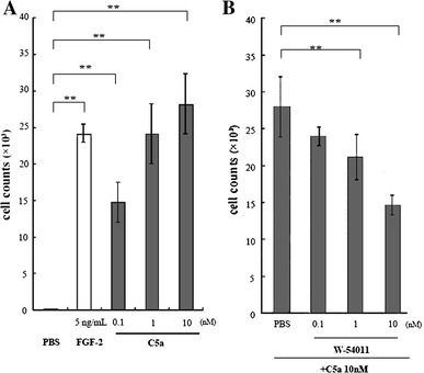
Analysis of HMEC-1 migration. HMEC-1 cells migrated to the reverse side of the filters were removed with trypsin and counted. a FGF-2 (5 ng/ml) was used as a positive control for inducing HMEC-1 migration (open column). C5a increased the migration of HMEC-1 cells in a dose-dependent manner (closed column), while this induced migration was inhibited by W-54011. b In a dose-dependent manner. Bars represent mean ± SEM (n = 3). a **P < 0.01 by Bonferroni’s multiple comparison test, b **P < 0.01 by Dunnett’s multiple comparison test
Fig. 7
Fig. 7
Endothelial cell infiltration and microvascular-like structures induced by C5a in vivo. Representative photomicrographs of Matrigel supplemented with phosphate-buffered saline (PBS) (a, e), 1,000 nM C3a (b, f), 1 μg/ml FGF-2 (c, g), or 100 nM C5a (d, h) stained with HE or anti-CD31 antibody. Endothelial cells stained positive with anti-CD31 antibody (black arrows) and these CD31+ cells formed microvascular-like structure (white arrows). Cell migration into the Matrigel was limited in the presence of PBS or C3a (a, e and b, f), whereas both FGF-2 and C5a had a significant effect on cell migration (c, g and d, h). Black arrows indicate CD31+ endothelial cells and white arrows indicate the microvascular-like structures formed by these cells in the presence of FGF-2 and C5a

Similar articles

Cited by

References

    1. Guo RF, Ward PA. Role of C5a in inflammatory responses. Annu Rev Immunol. 2005;23:821–852. doi: 10.1146/annurev.immunol.23.021704.115835. - DOI - PubMed
    1. Swerlick RA, Yancey KB, Lawley TJ. Inflammatory properties of human C5a and C5a des Arg/in mast cell-depleted human skin. J Invest Dermatol. 1989;93:417–422. doi: 10.1111/1523-1747.ep12280306. - DOI - PubMed
    1. Foreman KE, Vaporciyan AA, Bonish BK, Jones ML, Johnson KJ, Glovsky MM, et al. C5a-induced expression of P-selectin in endothelial cells. J Clin Invest. 1994;94:1147–1155. doi: 10.1172/JCI117430. - DOI - PMC - PubMed
    1. Watanabe M, Fujioka-Kaneko Y, Kobayashi H, Kiniwa M, Kuwano M, Basaki Y. Involvement of integrin-linked kinase in capillary/tube-like network formation of human vascular endothelial cells. Biol Proced Online. 2005;7:41–47. doi: 10.1251/bpo104. - DOI - PMC - PubMed
    1. Hirosumi J, Ouchi Y, Watanabe M, Kusunoki J, Nakamura T, Orimo H. Effects of growth factors on cytosolic free calcium concentration and DNA synthesis in cultured rat aortic smooth muscle cells. Tohoku J Exp Med. 1989;157:289–300. doi: 10.1620/tjem.157.289. - DOI - PubMed

Publication types

MeSH terms