

Molecular and cellular characterization of Neuregulin-1 type IV isoforms
- PMID: 20218976
- PMCID: PMC2931268
- DOI: 10.1111/j.1471-4159.2010.06677.x
Molecular and cellular characterization of Neuregulin-1 type IV isoforms
Abstract
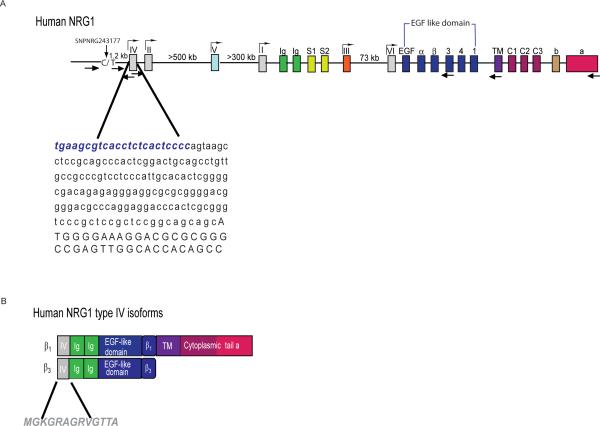
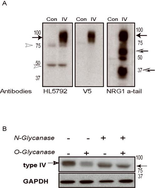
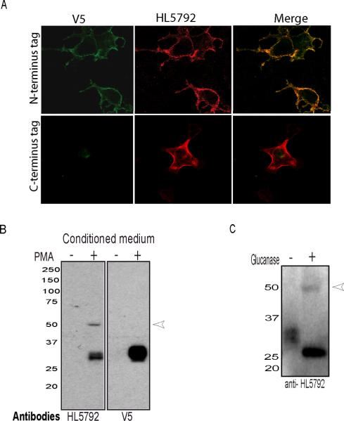
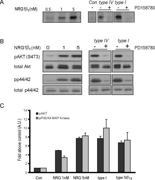
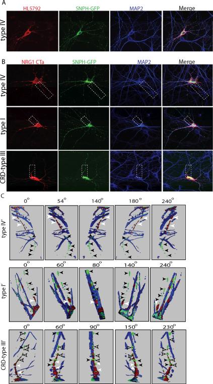
Numerous genetic studies associated the Neuregulin 1 (NRG1) Icelandic haplotype (HAP(ice)), and its single nucleotide polymorphism SNP8NRG243177 [T/T], with schizophrenia. Because SNP8NRG243177 [T/T] has characteristics of a functional polymorphism that maps close to NRG1 type IV coding sequences, our initial goal was to map precisely the human type IV transcription initiation site. We determined that the initiation site is 23 bp upstream of the previously reported type IV exon, and that no other transcripts map to the SNP8NRG243177 region. Because NRG1 type IV transcripts are specific to human, we isolated full-length NRG1 type IV cDNAs from human hippocampi and expressed them in non-neural cells and dissociated rat hippocampal neurons to study protein expression, processing and function. Using an antiserum we generated against the NRG1 type IV-specific N-terminus, we found that the protein is targeted to the cell surface where PKC activation promotes its cleavage and release of the extracellular domain. Conditioned medium derived from type IV expressing cells stimulates ErbB receptor phosphorylation, as well as downstream Akt and Erk signaling, demonstrating that NRG1 type IV possesses biological activity similar to other releasable NRG1 isoforms. To study the subcellular targeting of distinct isoforms, neurons were transfected with the Ig-domain-containing NRG1 types I and IV, or the cysteine-rich domain type III isoform. Three dimensional confocal images from transfected neurons indicate that, whereas all isoforms are expressed on somato-dendritic membranes, only the type III-cysteine-rich domain isoform is detectable in distal axons. These results suggest that NRG1 type IV expression levels associated with SNP8NRG243177 [T/T] can selectively modify signaling of NRG1 released from somato-dendritic compartments, in contrast to the type III NRG1 that is also associated with axons.
Figures
Similar articles
-
Molecular cloning of a brain-specific, developmentally regulated neuregulin 1 (NRG1) isoform and identification of a functional promoter variant associated with schizophrenia.J Biol Chem. 2007 Aug 17;282(33):24343-51. doi: 10.1074/jbc.M702953200. Epub 2007 Jun 12. J Biol Chem. 2007. PMID: 17565985
-
Structural Similarities between Neuregulin 1-3 Isoforms Determine Their Subcellular Distribution and Signaling Mode in Central Neurons.J Neurosci. 2017 May 24;37(21):5232-5249. doi: 10.1523/JNEUROSCI.2630-16.2017. Epub 2017 Apr 21. J Neurosci. 2017. PMID: 28432142 Free PMC article.
-
Specific regulation of NRG1 isoform expression by neuronal activity.J Neurosci. 2011 Jun 8;31(23):8491-501. doi: 10.1523/JNEUROSCI.5317-10.2011. J Neurosci. 2011. PMID: 21653853 Free PMC article.
-
Neuregulin 1 and schizophrenia: genetics, gene expression, and neurobiology.Biol Psychiatry. 2006 Jul 15;60(2):132-40. doi: 10.1016/j.biopsych.2005.11.002. Epub 2006 Jan 25. Biol Psychiatry. 2006. PMID: 16442083 Review.
-
The neuregulin-I/ErbB signaling system in development and disease.Adv Anat Embryol Cell Biol. 2007;190:1-65. Adv Anat Embryol Cell Biol. 2007. PMID: 17432114 Review.
Cited by
-
Neurobehavioral Differences Between Mice Receiving Distinct Neuregulin Variants as Neonates; Impact on Sensitivity to MK-801.Curr Mol Med. 2015;15(3):222-36. doi: 10.2174/1566524015666150330143300. Curr Mol Med. 2015. PMID: 25817857 Free PMC article.
-
Association study of neuregulin-1 gene polymorphisms in a North Indian schizophrenia sample.Schizophr Res. 2013 Mar;144(1-3):24-30. doi: 10.1016/j.schres.2012.12.017. Epub 2013 Jan 26. Schizophr Res. 2013. PMID: 23360725 Free PMC article.
-
Disruption of the neuregulin 1 gene in the rat alters HPA axis activity and behavioral responses to environmental stimuli.Physiol Behav. 2011 Aug 3;104(2):205-14. doi: 10.1016/j.physbeh.2010.11.015. Epub 2010 Nov 16. Physiol Behav. 2011. PMID: 21092742 Free PMC article.
-
Sex-specific neuroendocrine and behavioral phenotypes in hypomorphic Type II Neuregulin 1 rats.Behav Brain Res. 2011 Oct 31;224(2):223-32. doi: 10.1016/j.bbr.2011.05.008. Epub 2011 May 19. Behav Brain Res. 2011. PMID: 21620900 Free PMC article.
-
Schizophrenia-risk variant rs6994992 in the neuregulin-1 gene on brain developmental trajectories in typically developing children.Transl Psychiatry. 2014 May 27;4(5):e392. doi: 10.1038/tp.2014.41. Transl Psychiatry. 2014. PMID: 24865593 Free PMC article.
References
-
- Bjarnadottir M, Misner DL, Haverfield-Gross S, Bruun S, Helgason VG, Stefansson H, Sigmundsson A, Firth DR, Nielsen B, Stefansdottir R, Novak TJ, Stefansson K, Gurney ME, Andresson T. Neuregulin1 (NRG1) signaling through Fyn modulates NMDA receptor phosphorylation: differential synaptic function in NRG1+/− knock-outs compared with wild-type mice. J Neurosci. 2007;27:4519–4529. - PMC - PubMed
-
- Brewer GJ, Torricelli JR, Evege EK, Price PJ. Optimized survival of hippocampal neurons in B27-supplemented Neurobasal, a new serum-free medium combination. Journal of neuroscience research. 1993;35:567–576. - PubMed
-
- Buonanno A, Fischbach GD. Neuregulin and ErbB receptor signaling pathways in the nervous system. Curr Opin Neurobiol. 2001;11:287–296. - PubMed
-
- Collier DA, Li T. The genetics of schizophrenia: glutamate not dopamine? European journal of pharmacology. 2003;480:177–184. - PubMed
Publication types
MeSH terms
Substances
Grants and funding
LinkOut - more resources
Full Text Sources
Molecular Biology Databases
Research Materials
Miscellaneous
