. 2010 May;160(2):246-57.
doi: 10.1111/j.1476-5381.2010.00656.x. Epub 2010 Mar 9.

The metabolic enhancer piracetam ameliorates the impairment of mitochondrial function and neurite outgrowth induced by beta-amyloid peptide

Affiliations

The metabolic enhancer piracetam ameliorates the impairment of mitochondrial function and neurite outgrowth induced by beta-amyloid peptide

C Kurz et al. Br J Pharmacol. 2010 May.

Abstract

Background and purpose: beta-Amyloid peptide (Abeta) is implicated in the pathogenesis of Alzheimer's disease by initiating a cascade of events from mitochondrial dysfunction to neuronal death. The metabolic enhancer piracetam has been shown to improve mitochondrial dysfunction following brain aging and experimentally induced oxidative stress.

Experimental approach: We used cell lines (PC12 and HEK cells) and murine dissociated brain cells. The protective effects of piracetam in vitro and ex vivo on Abeta-induced impairment of mitochondrial function (as mitochondrial membrane potential and ATP production), on secretion of soluble Abeta and on neurite outgrowth in PC12 cells were investigated.

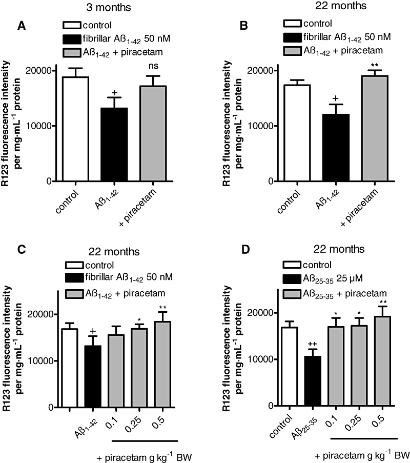
Key results: Piracetam improves mitochondrial function of PC12 cells and acutely dissociated brain cells from young NMRI mice following exposure to extracellular Abeta(1-42). Similar protective effects against Abeta(1-42) were observed in dissociated brain cells from aged NMRI mice, or mice transgenic for mutant human amyloid precursor protein (APP) treated with piracetam for 14 days. Soluble Abeta load was markedly diminished in the brain of those animals after treatment with piracetam. Abeta production by HEK cells stably transfected with mutant human APP was elevated by oxidative stress and this was reduced by piracetam. Impairment of neuritogenesis is an important consequence of Abeta-induced mitochondrial dysfunction and Abeta-induced reduction of neurite growth in PC12 cells was substantially improved by piracetam.

Conclusion and implications: Our findings strongly support the concept of improving mitochondrial function as an approach to ameliorate the detrimental effects of Abeta on brain function.


Figures

Figure 1
Figure 1
Piracetam, aniracetam, and oxiracetam ameliorate Aβ impaired mitochondrial function. (A) PC12 cells were treated for 24 h with fibrillar Aβ1-42, 30 min after insult piracetam (0.1 mM, 0.5 mM or 1 mM) was added and the MMP was measured using the fluorescence dye R123. Data are expressed as means ± SEM (n= 4–5). +P < 0.05 Aβ1-42 induced reduction of MMP versus untreated control; **P < 0.01, *P < 0.05 Aβ1-42 induced reduction of MMP versus piracetam treatment; student's unpaired t-test. (B) Dissociated brain cells isolated from NMRI mice were stressed for 4 h with fibrillar Aβ1-42. After 1 h, piracetam (0.1, 1, 10 mM) was added; again the mitochondrial membrane potential was detected. Data are expressed as means ± SEM (n= 4–5). +++P < 0.001 Aβ1-42 versus control; **P < 0.01 piracetam treatment versus Aβ1-42; student's unpaired t-test (C and D) PC12 cells were treated for 24 h with Aβ1-42, 30 min after insult oxiracetam (C) (0.5 mM or 1 mM), or aniracetam (D) (0.001 mM or 0.01 mM) were added and the MMP was investigated using R123. Data are expressed as means ± SEM (n= 4–5). +P < 0.05 Aβ1-42 versus control; **P < 0.01 oxiracetam or aniracetam treatment versus Aβ1-42, student's unpaired t-test (E) APPwt, APPsw and vctPC12 cells were stressed with SNP 0.5 mM in the presence and absence of piracetam (0.5 mM, 1 mM) and the MMP (E) and ATP levels (F) were measured. Data are presented as the reduction of MMP and ATP induced by SNP and the respective improvement by piracetam. PC12 cells were incubated 24 h with SNP, piracetam was added 30 min after insult. Data are expressed as means ± SEM (n= 4). *P < 0.05 SNP induced reduction of mitochondrial membrane potential in vct cells, APPwt, or APPsw cells versus vct cells, APPwt or APPsw cells treated with piracetam; *P < 0.05, **P < 0.01, ***P < 0.001 SNP induced reduction of ATP levels in vct cells, APPwt or APPsw cells versus ATP levels of vct cells, APPwt or APPsw cells treated with piracetam, student's unpaired t-test. APP, Amyloid precursor protein; APPsw, Swedish APP mutation; APPwt, Wildtype human APP; MMP, Mitochondrial membrane potential; R123, Rhodamine 123; SNP, Sodium nitroprusside.
Figure 2
Figure 2
Piracetam treatment improves MMP after Aβ1-42 induced stress in vitro and ex vivo. (A and B) Young NMRI mice (A, 3 months) and aged mice (B, 22 months) were treated with 0.5 g piracetam kg−1 in 0.9% NaCl solution p.o. once daily for 2 weeks. Dissociated brain cells were prepared and stressed with fibrillar Aβ1-42 (50 nM) for 4 h in the presence or absence of piracetam (1 mM). MMP was measured using R123. Data are expressed as means ± SEM (n= 6–8).+P < 0.05, ++P < 0.01 control against Aβ1-42 treated dissociated brain cells; ns; *P < 0.01 membrane potential of dissociated brain cells stressed with Aβ1-42 against dissociated brain cells treated with Aβ1-42 in the presence of piracetam; student's unpaired t-test. (C) Aged mice (22 months) received 0.1, 0.25, 0.5 g piracetam kg−1 in 0.9% NaCl solution p.o. once daily for 2 weeks. Control animals were treated with 0.9% NaCl solution alone. Afterwards, dissociated brain cells were incubated ex vivo for 4 h with 50 nM fibrillar Aβ1-42 and MMP was measured. Piracetam protects against Aβ1-42 and Aβ25-35 induced mitochondrial damage ex vivo. Membrane potential of dissociated brain cells was measured after 4 h incubation with 50 nM Aβ1-42 (C) and 25 µM Aβ25-35 (D). Data are expressed as means ± SEM (n= 6–8) +P < 0.05, ++P < 0.01 untreated control versus Aβ1-42 induced reduction of MMP; *P < 0.05, **P < 0.01 Aβ1-42 or Aβ25-35 reduced MMP versus piracetam treated groups; student's unpaired t-test. MMP, Mitochondrial membrane potential; R123, Rhodamine 123.
Figure 3
Figure 3
Piracetam improves mitochondrial function in tgAPP mice and reduces Aβ1-40 levels in tgAPP mice and APPwt HEK290 cells. Treated animals received 0.5 g piracetam kg−1 in 0.9% NaCl solution p.o. once daily for 2 weeks. Control animals were treated with 0.9% NaCl solution alone. (A) The MMP was significantly reduced in tgAPP mice. Piracetam treatment normalizes the MMP to non-tgAPP levels. Data are expressed as means ± SEM (n= 7–8). +P < 0.05 control non-tgAPP versus control tgAPP, *P < 0.01 piracetam treated tgAPP versus tgAPP control; student's unpaired t-test. (B) ATP levels were also impaired in tgAPP mice. In contrast, piracetam treatment increases ATP levels not only in tgAPP animals but also in control animals. Data are expressed as means ± SEM (n= 7–8). *P < 0.05 control non-tgAPP versus piracetam treated non-tgAPP; +P < 0.05 control non-tgAPP versus control tgAPP, *P < 0.01 piracetam treated tgAPP versus tgAPP control; student's unpaired t-test. (C) Piracetam reduced Aβ1-40 levels in tg-APP mice. Normalized Aβ levels were quantified with elisa in Tris-buffered brain homogenates from non-tg littermate and tgAPP mice (3 months old). Data are expressed as means ± SEM (n= 7–8). +++P < 0.05 control non-tgAPP versus control tgAPP, **P < 0.01 piracetam treated tgAPP versus control tgAPP; student's unpaired t-test. (D) Piracetam reduced Aβ levels in APPwt HEK293 cells. Cells were incubated for 24 h with piracetam (1 mM) and Aβ1-40 levels were investigated using the Aβ1-40 elisa. Data are expressed as means ± SEM (n= 3–4). **P < 0.01 APPwt HEK293 control cells versus cells treated with piracetam 1 mM; student's unpaired t-test. (E) Piracetam improved MMP under basal conditions in APPwt HEK293 cells. Cells were incubated for 24 h with piracetam (1 mM) and MMP was measured using R123. Data are expressed as means ± SEM (n= 6). ***P < 0.001 APPwt HEK293 control cells versus cells treated with piracetam; student's unpaired t-test. (F) Piracetam reduced nitrosative stress-induced elevation of Aβ in APPwt HEK293 cells. Cells were pre-incubated for 24 h with piracetam (1 mM) and stressed for additional 24 h with SNP (0.5 mM). Aβ levels were again detected using the Aβ1-40 elisa. Data are expressed as means ± SEM (n= 3–4). +++P < 0.001 APPwt HEK293 control cells versus cells treated with SNP, *P < 0.01 APPwt HEK293 cells stressed with SNP 0.5 mM versus stressed cells pre-incubated with piracetam; student's unpaired t-test. (G) Piracetam ameliorated nitrosative stress-induced reduction of MMP. Cells were pre-incubated for 24 h with piracetam (1 mM) and stressed for additional 24 h with SNP (0.5 mM). Data are expressed as means ± SEM (n= 6). **P < 0.01 APPwt HEK293 cells stressed with SNP 0.5 mM versus stressed cells pre-incubated with piracetam, student's unpaired t-test. Aβ, β-Amyloid; APPwt, Wildtype human amyloid precursor protein; MMP, Mitochondrial membrane potential; tgAPP, Transgenic for the human amyloid precursor protein.
Figure 5
Figure 5
Piracetam improves the effect of the neurotrophin NGF in APPwt and APPsw PC12 cells on neurite outgrowth. APPwt, APPsw and vct PC12 cells were treated over 6 days with NGF 50 ng·mL−1 in the presence or absence of piracetam 1 mM and neurite outgrowth was measured. (A) The neurite length in APPsw PC12 cells was significantly reduced compared to vct PC12 cells (transfected with the corresponding vector). (B) In the presence of piracetam the neurite length after treatment with NGF is improved in all three cell types. The effect is normalized to the respective control (without piracetam, 100%). (C) In the presence of different NGF concentrations (1–50 ng·mL−1) piracetam improved the neurotrophic effect of NGF in APPwt (C) and APPsw cells (D). Data are expressed as means ± SEM (n= 6–7) **P < 0.01, ***P < 0.001 versus the respective control, two-way anova with Bonferroni's post test. APPsw, Swedish amyloid precursor protein mutation; APPwt, Wildtype human amyloid precursor protein; NGF, Nerve growth factor.
Figure 4
Figure 4
Piracetam protects against Aβ and SNP induced impairment of neurite outgrowth. PC12 cells were treated over 6 days with NGF 50 ng·mL−1 in the presence or absence of oligomeric Aβ 1 µM or SNP 0.05 mM. In addition, piracetam 1 mM was added and the effects on neurite outgrowth were investigated. (A) Representative images from cells treated with NGF 50 ng·mL−1, NGF + oligomeric Aβ, and NGF + oligomeric Aβ and piracetam. (B) Neurite length of PC12 cells treated with NGF, NGF + oligomeric Aβ, and NGF + oligomeric Aβ. Data are expressed as means ± SEM (n= 6–7) ++P < 0.01 NGF versus oligomeric Aβ; ++P < 0.01 NGF + oligomeric Aβ versus NGF + oligomeric Aβ+ piracetam, student's unpaired t-test. (C) Neurite length of PC12 cells treated with NGF, NGF + SNP 0.05 mM, NGF + SNP 0.05 mM, NGF + SNP 0.05 mM + piracetam. Data are expressed as means ± SEM (n= 6–7) +++P < 0.001 NGF versus NGF + SNP; *P < 0.05 NGF + SNP 0.5 mM versus NGF + SNP 0.05 mM + piracetam, student's unpaired t-test. Aβ, β-Amyloid; NGF, Nerve growth factor; SNP, Sodium nitroprusside.

Comment in

Similar articles

Cited by

References

    1. Alexander SPH, Mathie A, Peters JA. Guide to Receptors and Channels (GRAC), 4th edition. Br J Pharmacol. 2009;158(Suppl. 1):S1–S254. - PMC - PubMed
    1. Atamna H, Nguyen A, Schultz C, Boyle K, Newberry J, Kato H, et al. Methylene blue delays cellular senescence and enhances key mitochondrial biochemical pathways. FASEB J. 2008;22:703–712. - PubMed
    1. Bachurin SA, Shevtsova EP, Kireeva EG, Oxebkrug GF, Sablin SO. Mitochondria as a target for neurotoxins and neuroprotective agents. Ann N Y Acad Sci. 2003;993:334–344. - PubMed
    1. Benzi G, Pastoris O, Villa RR, Giuffrida AM. Influence of aging and exogenous substances on cerebral energy metabolism in posthypoglycemic recovery. Biochem Pharmacol. 1985;34:1477–1483. - PubMed
    1. Bernales S, Wagner S, Protter AA, Hung DT. Dimebon induces neurite outgrowth and mitochondrial statilizion. Washington, DC: Society for Neuroscience; 2008. Program No. 543.29/S12. Neuroscience Meeting Planner. Online.

Publication types