. 2010 Mar 10:9:56.
doi: 10.1186/1476-4598-9-56.

Specific effects of bortezomib against experimental malignant pleural effusion: a preclinical study

Affiliations

Specific effects of bortezomib against experimental malignant pleural effusion: a preclinical study

Ioannis Psallidas et al. Mol Cancer. .

Abstract

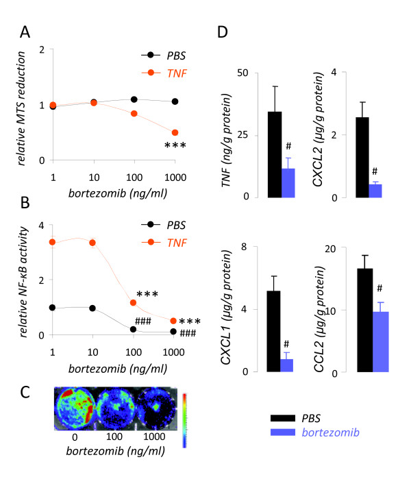
Background: We have previously shown that nuclear factor (NF)-kappaB activation of mouse Lewis lung carcinoma (LLC) specifically promotes the induction of malignant pleural effusions (MPE) by these cells. In the present studies we hypothesized that treatment of immunocompetent mice with bortezomib tailored to inhibit cancer cell NF-kappaB activation and not proliferation specifically inhibits MPE formation by LLC cells.

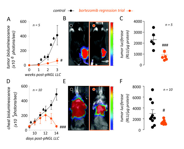
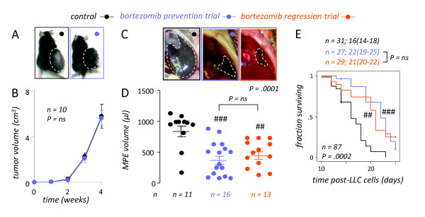
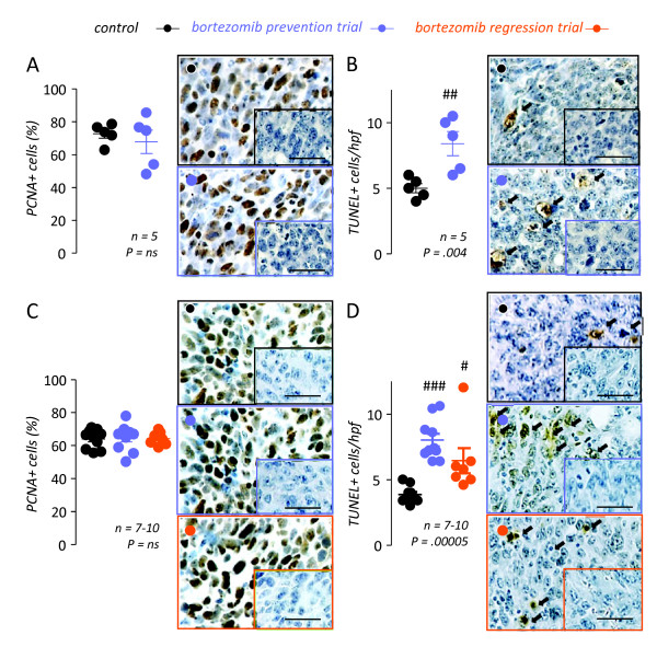
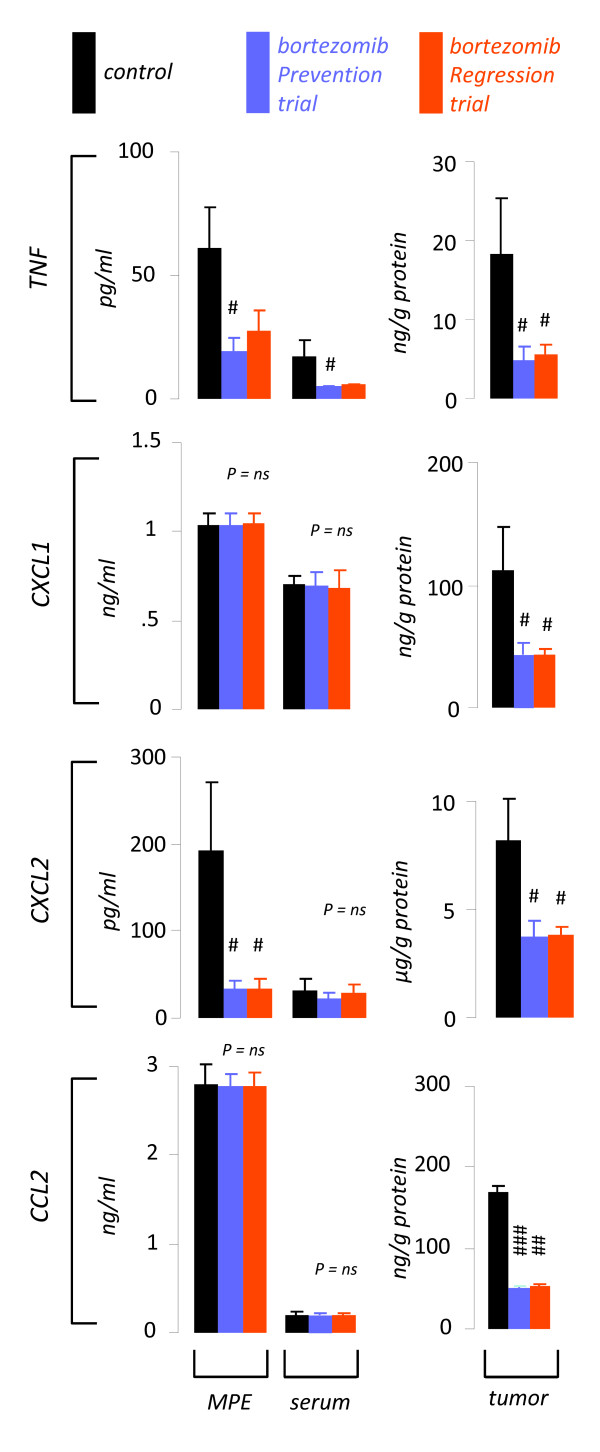
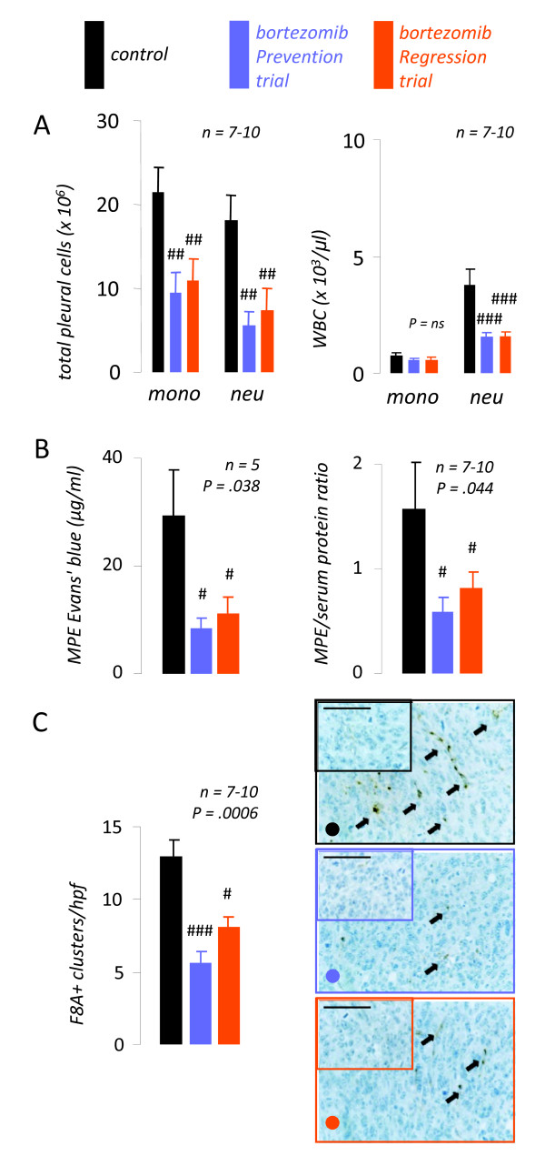
Results: Treatment of LLC cells with low concentrations of bortezomib (100 ng/ml) inhibited NF-kappaB activation and NF-kappaB-dependent transcription, but not cellular proliferation. Bortezomib treatment of immunocompetent C57BL/6 mice bearing LLC-induced subcutaneous tumors and MPEs significantly blocked tumor-specific NF-kappaB activation. However, bortezomib treatment did not impair subcutaneous LLC tumor growth, but was effective in limiting LLC-induced MPE. This specific effect was evidenced by significant reductions in effusion accumulation and the associated mortality and was observed with both preventive (beginning before MPE formation) and therapeutic (beginning after MPE establishment) bortezomib treatment. The favorable impact of bortezomib on MPE was associated with suppression of cardinal MPE-associated phenomena, such as inflammation, vascular hyperpermeability, and angiogenesis. In this regard, therapeutic bortezomib treatment had identical favorable results on MPE compared with preventive treatment, indicating that the drug specifically counteracts effusion formation.

Conclusions: These studies indicate that proteasome inhibition tailored to block NF-kappaB activation of lung adenocarcinoma specifically targets the effusion-inducing phenotype of this tumor. Although the drug has limited activity against advanced solid lung cancer, it may prove beneficial for patients with MPE.


Figures

Figure 1
Figure 1
Tailoring of bortezomib treatment to target nuclear factor (NF)-κB activation of mouse lung adenocarcinoma cells in vitro. (A) Relative MTS reduction capacity of Lewis lung cancer (LLC) cells after 24 hours of incubation with or without 1 nM recombinant human (rh) TNF in the presence of varying concentrations of bortezomib. (B) Relative nuclear factor (NF)-κB-dependent luciferase expression of NF-κB. GFP.luc (pNGL) LLC cells after 4 hours of incubation with or without 1 nM rh TNF in the presence of varying concentrations of bortezomib. (C) Bioluminescence image of pNGL LLC cells exposed for 4 hours to the indicated doses of bortezomib. Colors on the top of the scale indicate high light emission. (D) Cytokine/chemokine release by wild-type LLC cells incubated with or without 100 ng/ml bortezomib for 24 hours. TNF, tumor necrosis factor; CXCL, C-X-C motif chemokine ligand; CCL, C-C motif chemokine ligand. All experiments were done thrice. Columns, dots, mean; bars, standard error of mean. # and ###: P < .05 and .001, respectively, compared with cells treated with PBS. * and ***: P < .05 and .001, respectively, compared with cells treated with 1 nM rhTNF.
Figure 2
Figure 2
Tailored bortezomib treatment inhibits tumor-specific nuclear factor (NF)-κB activation of subcutaneous and intrapleural lung adenocarcinoma. (A-C) NF-κB-targeted bortezomib treatment blocks NF-κB activation of subcutaneous adenocarcinoma in vivo. Wild-type C57BL/6 mice received 5 × 10⁵ subcutaneous NF-κB-reporter (NF-κB. GFP.luc, pNGL) Lewis lung carcinoma (LLC) cells and were allowed one week for tumors to implant, where after they received bi-weekly intraperitoneal bortezomib (100 ng/g = 0.1 mg/kg) or PBS in a regression trial. Mice were serially imaged for bioluminescence after intravenous injection of 1 mg D-luciferin at the time-points indicated (A, B) and were sacrificed on day 21 for determination of NF-κB-dependent luciferase bioactivity of tumor tissue homogenates (C). Shown are time course of tumor-specific NF-κB activity in flank tumors (A), representative bioluminescence images at day 21 (B), and summary of luciferase assay data obtained at day 21 (C). (D-F) NF-κB-targeted bortezomib treatment blocks NF-κB activation of intrapleural adenocarcinoma in vivo. C57BL/6 mice received 1.5 × 10⁵ intrapleural pNGL LLC cells and were allowed one week, where after they received bi-weekly bortezomib (100 ng/g = 0.1 mg/kg) or PBS. Mice were imaged for bioluminescence (D, E) and were sacrificed on day 14 for determination of NF-κB-dependent luciferase bioactivity of tumor tissue (F). Shown are time course of tumor-specific NF-κB activity in MPE-bearing mice (D), representative bioluminescence images at day 14 (E), and summary of luciferase assay data obtained at day 14 (F). n, sample size; RLU, relative light units. Dots, mean (left) or raw data points (right); lines in panels on the right, mean; bars, standard error of mean. # and ###: P < .05 and .001, respectively, compared with control.
Figure 3
Figure 3
NF-κB-targeted bortezomib treatment specifically inhibits MPE but not subcutaneous tumor formation by lung adenocarcinoma. (A, B) NF-κB-targeted bortezomib treatment has no effect on subcutaneous Lewis lung carcinoma (LLC) growth. C57BL/6 mice received 5 × 10⁵ subcutaneous LLC cells followed by bi-weekly intraperitoneal bortezomib (100 ng/g = 0.1 mg/kg) or PBS. Separate subsets of mice received treatment either immediately (prevention trial) or starting one week after tumor implant (regression trial). Tumor volume was determined weekly and mice were sacrificed on day 28. Representative images at 4 weeks (A; dashed lines outline tumors) and mean volume (B) of subcutaneous tumors in PBS and bortezomib treated mice. Results from prevention and regression trial were identical and were grouped for demonstration purposes. Dots, mean; bars, standard error of mean. (C-E) NF-κB-targeted bortezomib treatment is effective against malignant pleural effusion (MPE) induced by LLC cells. C57BL/6 mice received 1.5 × 10⁵ intrapleural LLC cells followed by bi-weekly bortezomib (100 ng/g = 0.1 mg/kg) or PBS. Mice were enrolled in prevention (immediate treatment) or regression (treatment starting after one week) trials and were sacrificed on day 14 (C, D) or observed till moribund (E). (C) Representative transdiaphragmatic photographs at 14 days (dashed lines outline MPEs) and (D) mean volume of MPE in PBS- and bortezomib-treated mice. Dots, raw data points; lines, mean; bars, SEM. (E) Survival of MPE-bearing mice treated with PBS or bortezomib. Shown are Kaplan-Meier survival curves and estimates (median, 95% confidence interval) of pooled data from three independent experiments. (D, E) Note that results from prevention and regression trials were not significantly different (ns). n, sample size; P, probability values. ## and ###: P < .01 and .001, respectively, compared with control.
Figure 4
Figure 4
NF-κB-targeted bortezomib treatment does not impact tumor cell proliferation in vivo. Subcutaneous (A, B) and pleural (C, D) tumor tissue was obtained on days 28 and 14, respectively, from experiments described in Figure 3 and was immunostained for proliferating cell nuclear antigen (PCNA; A, C) and terminal deoxynucleotidyl nick-end labeling (TUNEL; B, D), indicators of cell proliferation and apoptosis, respectively. Shown are summary of results and representative photomicrographs (Å = 600; scale bars = 50 μm). NF-κB-targeted bortezomib treatment had no effect on Lewis lung carcinoma (LLC) cell proliferation in subcutaneous or pleural tumors (A, C). However, bortezomib treatment resulted in enhanced apoptosis rates in both tumor sites (B, D). n, sample size; P, probability values; hpf, high-power field. Dots, raw data points; lines, mean; bars, standard error of mean. #, ##, and ###: P < .05, .01, and .001, respectively, compared with control.
Figure 5
Figure 5
Tailored bortezomib treatment down-regulates NF-κB-dependent paracrine mediator expression of pleural lung adenocarcinoma. Pleural fluid, blood, and pleural tumor tissue NF-κB-dependent gene product levels were determined in samples obtained at day 14 from mice with MPE enrolled in control (n = 10), bortezomib prevention (n = 10), and bortezomib regression (n = 7) trials as in Figure 3C-D. TNF, tumor necrosis factor; CXCL, C-X-C motif chemokine ligand; CCL, C-C motif chemokine ligand. Columns, mean; bars, standard error of mean; n, sample size; P, overall probability values. #, ## and ###: P < .05, .01, and .001, respectively, compared with control.
Figure 6
Figure 6
NF-κB-targeted bortezomib inhibits malignant pleural effusion (MPE)-associated inflammation, vascular leakiness, and angiogenesis. Mice were treated as in Figure 3C-D. (A) Pleural fluid and blood inflammatory cell numbers at day 14. (B) Left: MPE Evans' blue levels determined at day 14, one hour after intravenous delivery of 0.8 mg of the dye. Right: MPE/serum protein ratio. Both Evans' blue leakage and protein ration are indicators of vascular hyperpermeability. (C) Pleural tumor tissue microvessel density assessed by immunoreactivity for factor VIII-associated protein (F8A). Summary of data (left) and representative microphotographs (right; Å = 400; scale bar = 100 μm; arrows, new vessels). MPE, malignant pleural effusion; WBC, white blood cells; mono, mononuclear cells; neu, neutrophils; hpf, high-power field. Columns, mean; bars, standard error of mean; n, sample size; P, overall probability values. #, ## and ###: P < .05, .01, and .001, respectively, compared with control.

Similar articles

Cited by

References

    1. Antony VB, Loddenkemper R, Astoul P, Boutin C, Goldstraw P, Hott J, Rodriguez Panadero F, Sahn SA. Management of malignant pleural effusions. Eur Respir J. 2001;18:402–419. doi: 10.1183/09031936.01.00225601. - DOI - PubMed
    1. Antunes G, Neville E, Duffy J, Ali N. Pleural Diseases Group, Standards of Care Committee, British Thoracic Society. BTS guidelines for the management of malignant pleural effusions. Thorax. 2003;58(Suppl 2):ii29–38. - PMC - PubMed
    1. Sugiura S, Ando Y, Minami H, Ando M, Sakai S, Shimokata K. Prognostic value of pleural effusion in patients with non-small cell lung cancer. Clin Cancer Res. 1997;3:47–50. - PubMed
    1. Tremblay A. Tunnelled pleural catheters in malignant pleural effusion. Lancet. 2007;370:387. doi: 10.1016/S0140-6736(07)61189-1. - DOI - PubMed
    1. Viallat JR, Rey F, Astoul P, Boutin C. Thoracoscopic talc poudrage pleurodesis for malignant effusions. A review of 360 cases. Chest. 1996;110:1387–1393. doi: 10.1378/chest.110.6.1387. - DOI - PubMed

Publication types

MeSH terms

LinkOut - more resources