

Sodium valproate-induced congenital cardiac abnormalities in mice are associated with the inhibition of histone deacetylase
- PMID: 20219112
- PMCID: PMC2841099
- DOI: 10.1186/1423-0127-17-16
Sodium valproate-induced congenital cardiac abnormalities in mice are associated with the inhibition of histone deacetylase
Abstract
Background: Valproic acid, a widely used anticonvulsant drug, is a potent teratogen resulting in various congenital abnormalities. However, the mechanisms underlying valproic acid induced teratogenesis are nor clear. Recent studies indicate that histone deacetylase is a direct target of valproic acid.
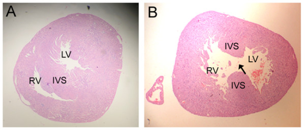
Methods: In the present study, we have used histological analysis and RT-PCR assays to examine the cardiac abnormalities in mice treated with sodium valproate (NaVP) and determined the effects of NaVP on histone deacetylase activity and the expression of heart development-related genes in mouse myocardial cells.
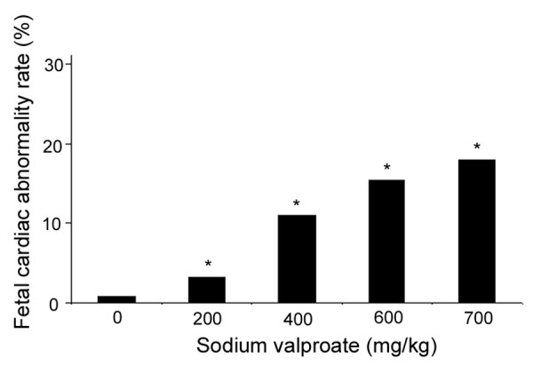
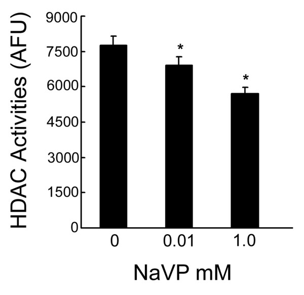
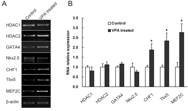
Results: The experimental data show that NaVP can induce cardiac abnormalities in fetal mice in a dose-dependent manner. NaVP causes a dose-dependent inhibition of hitone deacetylase (HDAC) activity in mouse myocardial cells. However, the expression levels of HDAC (both HDAC1 and HDAC2) are not significantly changed in fetal mouse hearts after administration of NaVP in pregnant mice. The transcriptional levels of other heart development-related genes, such as CHF1, Tbx5 and MEF2, are significantly increased in fetal mouse hearts treated with NaVP.
Conclusions: The study indicates that administration of NaVP in pregnant mice can result in various cardiac abnormalities in fetal hearts, which is associated with an inhibition of histone deacetylase without altering the transcription of this enzyme.
Figures
Similar articles
-
Disruption of Planar Cell Polarity Pathway Attributable to Valproic Acid-Induced Congenital Heart Disease through Hdac3 Participation in Mice.Chin Med J (Engl). 2018 Sep 5;131(17):2080-2088. doi: 10.4103/0366-6999.239311. Chin Med J (Engl). 2018. PMID: 30127218 Free PMC article.
-
Histone deacetylase is a direct target of valproic acid, a potent anticonvulsant, mood stabilizer, and teratogen.J Biol Chem. 2001 Sep 28;276(39):36734-41. doi: 10.1074/jbc.M101287200. Epub 2001 Jul 25. J Biol Chem. 2001. PMID: 11473107
-
Inhibition of histone deacetylases protects septic mice from lung and splenic apoptosis.J Surg Res. 2014 Apr;187(2):559-70. doi: 10.1016/j.jss.2013.10.050. Epub 2013 Oct 29. J Surg Res. 2014. PMID: 24290430
-
Valproic acid in pregnancy: how much are we endangering the embryo and fetus?Reprod Toxicol. 2009 Jul;28(1):1-10. doi: 10.1016/j.reprotox.2009.02.014. Epub 2009 Mar 13. Reprod Toxicol. 2009. PMID: 19490988 Review.
-
Mode of action: inhibition of histone deacetylase, altering WNT-dependent gene expression, and regulation of beta-catenin--developmental effects of valproic acid.Crit Rev Toxicol. 2005 Oct-Nov;35(8-9):727-38. doi: 10.1080/10408440591007403. Crit Rev Toxicol. 2005. PMID: 16417040 Review.
Cited by
-
[Effect of histone acetylation/deacetylation imbalances on key gene of planar cell polarity pathway].Zhongguo Dang Dai Er Ke Za Zhi. 2017 Apr;19(4):475-483. doi: 10.7499/j.issn.1008-8830.2017.04.022. Zhongguo Dang Dai Er Ke Za Zhi. 2017. PMID: 28407839 Free PMC article. Chinese.
-
Protein acetylation and deacetylation: An important regulatory modification in gene transcription (Review).Exp Ther Med. 2020 Oct;20(4):2923-2940. doi: 10.3892/etm.2020.9073. Epub 2020 Jul 29. Exp Ther Med. 2020. PMID: 32855658 Free PMC article. Review.
-
Chromatin modifying agents in the in vitro production of bovine embryos.Vet Med Int. 2010 Sep 29;2011:694817. doi: 10.4061/2011/694817. Vet Med Int. 2010. PMID: 20936105 Free PMC article.
-
Transcriptome-based prediction of drugs, inhibiting cardiomyogenesis in human induced pluripotent stem cells.Cell Death Discov. 2023 Aug 29;9(1):321. doi: 10.1038/s41420-023-01616-6. Cell Death Discov. 2023. PMID: 37644023 Free PMC article.
-
Prenatal S-Adenosine Methionine (SAMe) Induces Changes in Gene Expression in the Brain of Newborn Mice That Are Prevented by Co-Administration of Valproic Acid (VPA).Int J Mol Sci. 2020 Apr 18;21(8):2834. doi: 10.3390/ijms21082834. Int J Mol Sci. 2020. PMID: 32325788 Free PMC article.
References
Publication types
MeSH terms
Substances
Grants and funding
LinkOut - more resources
Full Text Sources
Medical
Miscellaneous
