. 2010 Mar 10:8:23.
doi: 10.1186/1477-7827-8-23.

Effect of adiponectin on bovine granulosa cell steroidogenesis, oocyte maturation and embryo development

Affiliations

Effect of adiponectin on bovine granulosa cell steroidogenesis, oocyte maturation and embryo development

Virginie Maillard et al. Reprod Biol Endocrinol. .

Abstract

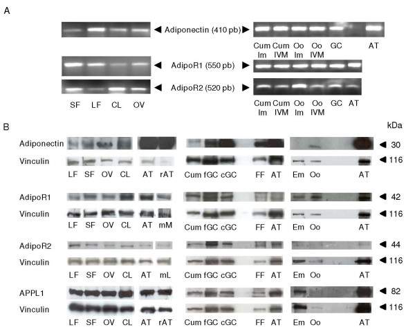
Background: Adiponectin is an adipokine, mainly produced by adipose tissue. It regulates several reproductive processes. The protein expression of the adiponectin system (adiponectin, its receptors, AdipoR1 and AdipoR2 and the APPL1 adaptor) in bovine ovary and its role on ovarian cells and embryo, remain however to be determined.

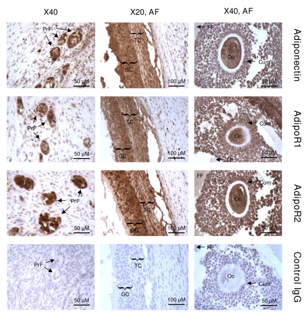
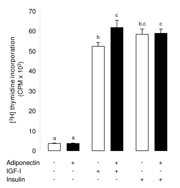
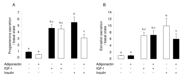
Methods: Here, we identified the adiponectin system in bovine ovarian cells and embryo using RT-PCR, immunoblotting and immunohistochemistry. Furthermore, we investigated in vitro the effects of recombinant human adiponectin (10 micro g/mL) on proliferation of granulosa cells (GC) measured by [3H] thymidine incorporation, progesterone and estradiol secretions measured by radioimmunoassay in the culture medium of GC, nuclear oocyte maturation and early embryo development.

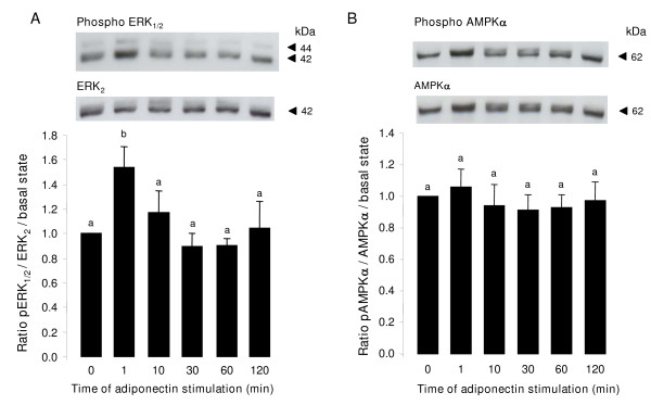
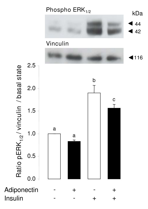
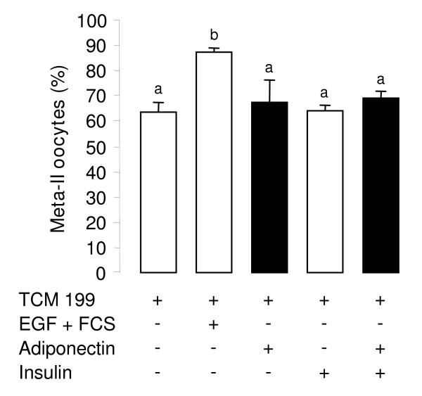
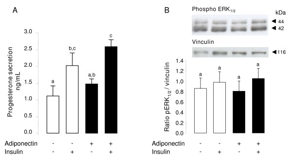
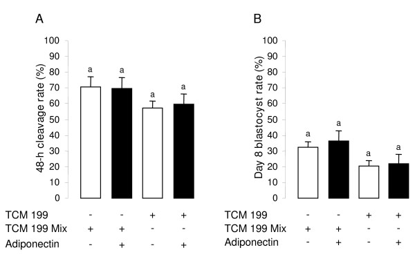
Results: We show that the mRNAs and proteins for the adiponectin system are present in bovine ovary (small and large follicles and corpus luteum) and embryo. Adiponectin, AdipoR1 and AdipoR2 were more precisely localized in oocyte, GC and theca cells. Adiponectin increased IGF-1 10(-8) M-induced GC proliferation (P < 0.01) but not basal or insulin 10(-8) M-induced proliferation. Additionally, adiponectin decreased insulin 10(-8) M-induced, but not basal or IGF-1 10(-8) M-induced secretions of progesterone (P < 0.01) and estradiol (P < 0.05) by GC. This decrease in insulin-induced steroidogenesis was associated with a decrease in ERK1/2 MAPK phosphorylation in GC pre-treated with adiponectin. Finally, addition of adiponectin during in vitro maturation affected neither the percentage of oocyte in metaphase-II nor 48-h cleavage and blastocyst day 8 rates.

Conclusions: In bovine species, adiponectin decreased insulin-induced steroidogenesis and increased IGF-1-induced proliferation of cultured GC through a potential involvement of ERK1/2 MAPK pathway, whereas it did not modify oocyte maturation and embryo development in vitro.


Figures

Figure 1
Figure 1
Expression of adiponectin system (mRNA and proteins) in bovine ovary and embryo. (A) RT-PCR analysis of the mRNAs for adiponectin, AdipoR1 and AdipoR2 in small (SF) and large (LF) follicles, corpus luteum (CL), whole ovary (Ov), cumulus cells from immature (Cum Im) and 24-h IVM (Cum IVM) COC, immature (Oo Im) and 24-h IVM (Oo IVM) oocytes, granulosa cells (GC) and adipose tissue (AT). (B) Detection of adiponectin (30 kDa), AdipoR1 (42 kDa), AdipoR2 (44 kDa) and APPL1 (82 kDa) by immunoblotting in LF, SF, OV, CL, AT, cumulus cells (Cum, from 40 24-h IVM COC per lane), fresh (fGC) and 48-h cultured (cGC) granulosa cells from small follicles (< 6 mm), follicular fluid (FF), oocytes (Oo, n = 100 per lane; except for APPL1, n = 20 per lane) and embryo (Em, in vitro day-8 blastocyst, n = 55 per lane). Rat adipose tissue (rAT) was used as positive control for the presence of adiponectin and APPL1, as already tested in our laboratory with the present antibodies. Mouse muscle (mM) and mouse liver (mL) were used as positive controls for the presence of AdipoR1 and AdipoR2 respectively, because these two antibodies were described to cross react with mouse. Vinculin protein was used as a loading control (n = 3).
Figure 2
Figure 2
Localization of adiponectin, AdipoR1 and AdipoR2 in bovine ovary by immunohistochemistry. Adiponectin, AdipoR1 and AdipoR2 were detected in primary (PrF) and antral (AF) follicles, granulosa cells (GC), cumulus cells (Cum), theca cells (TC), oocyte (Oo), and follicular fluid (FF). Negative controls included a section incubated with rabbit IgG (n = 3).
Figure 3
Figure 3
Effect of rh adiponectin on proliferation of bovine granulosa cells. [3H] thymidine incorporation was determined in bovine granulosa cells cultured for 24 h in enriched McCoy's 5A medium (without FBS) supplemented with or without rh adiponectin (10 μg/mL), ± IGF-I (10-8 M), or insulin (10-8 M). The data are expressed as mean ± SE and the measure unit of [3H] thymidine incorporation is counts per min (CPM). The results are representative of three independent cultures with each condition in triplicate. Bars with different superscripts are significantly different (P < 0.05).
Figure 4
Figure 4
Effect of rh adiponectin on steroidogenesis of bovine granulosa cells. Progesterone (A) and estradiol (B) secretions were measured by RIA protocol in culture medium of granulosa cells after 48 h of culture in enriched McCoy's 5A medium (without FBS) with or without rh adiponectin (10 μg/mL), +/- IGF-I (10-8 M), or insulin (10-8 M). The data are expressed as the amount of steroids (ng/mL) secreted per 48 h per 50 μg protein and per basal amount. The results, expressed as means ± SE, are representative of four independent cultures with each condition in quintuplet. Bars with different superscripts are significantly different (P < 0.05).
Figure 5
Figure 5
Effect of rh adiponectin on phosphorylation of MAPK ERK1/2 (A) and AMPK (B) in bovine granulosa cells. Granulosa cells from small follicles were cultured for 24 h in enriched McCoy's 5A medium supplemented with 10% FBS, starved of serum for 18 h and then incubated in serum-free medium with or without adiponectin (10 μg/mL) from 1 to 120 min. Protein extracts were separated by electrophoresis on 12% (w:v) SDS-polyacrylamide gel. After transfer to nitrocellulose membranes, the proteins were probed with anti-phospho-ERK1/2 or anti-phospho-AMPKα. The blots were stripped and reprobed with antibodies against ERK2 or AMPKα, respectively. Bands on the blots were quantified and the phosphorylated/total protein ratio was calculated. Values represent means ± SE relative to the basal state from at least three independent experiments. Bars with different superscripts are significantly different (P < 0.05).
Figure 6
Figure 6
Effect of insulin after 24-h stimulation with rh adiponectin on phosphorylation of MAPK ERK1/2 in bovine granulosa cells. Granulosa cells from small follicles were cultured for 24 h in enriched McCoy's 5A medium supplemented with 10% FBS, starved of serum for 24 h and then incubated in serum-free medium with or without rh adiponectin (10 μg/mL) for 24 h. Then, granulosa cells were cultured in fresh serum-free medium with or without insulin (10-8 M) for 5 min. Protein extracts were separated by electrophoresis on 12% (w:v) SDS-polyacrylamide gel. After transfer to nitrocellulose membranes, the proteins were probed with anti-phospho-ERK1/2. The blots were stripped and reprobed with antibodies against vinculin. Bands on the blots were quantified and the phospho-ERK1/2/vinculin ratio was calculated. Values represent means ± SE relative to the basal state from three independent experiments. Bars with different superscripts are significantly different (P < 0.05).
Figure 7
Figure 7
Effect of rh adiponectin on 24-h nuclear maturation of bovine oocyte in vitro. In vitro maturation of COC was performed in TCM 199 serum-free medium (control) supplemented or not with EGF (10 ng/mL) + 10% FBS (positive control), or rh adiponectin (10 μg/mL) ± insulin (10-8 M) for 24 h. The percentage of oocytes in metaphase-II stage was determined by chromatin labelling with Hoechst33342, as described in material and methods section. The data represents mean values ± SE from three independent experiments. Bars with different superscripts are significantly different (P < 0.05).
Figure 8
Figure 8
Effect of rh adiponectin on progesterone secretion by bovine COC (A) and phosphorylation of MAPK ERK1/2 in these complexes (B). Progesterone secretion (A) was measured by RIA protocol in culture medium of COC (20 COC/200 μL medium/well) after 24 h of IVM in TCM 199 serum-free medium supplemented or not with rh adiponectin (10 μg/mL) +/- insulin (10-8 M). The data are expressed as the amount of progesterone (ng/mL) secreted per 24 h. The results, expressed as means ± SE, are representative of four independent cultures. (B) IVM of COC (15 COC/well) was performed in TCM 199 serum-free medium supplemented or not with rh adiponectin (10 μg/mL) ± insulin (10-8 M) for 1 h. Protein extracts were separated by electrophoresis on 12% (w:v) SDS-polyacrylamide gel. After transfer to nitrocellulose membranes, the proteins were probed with anti-phospho-ERK1/2. The blots were stripped and reprobed with antibodies against vinculin. Bands on the blots were quantified and the phospho-ERK1/2/vinculin ratio was calculated. Values represent means ± SE relative to the basal state from three independent experiments. Bars with different superscripts are significantly different (P < 0.05).
Figure 9
Figure 9
Effect of rh adiponectin on bovine early embryo development in vitro. After 24 h of IVM in TCM 199 serum-free medium or TCM 199 mix with or without rh adiponectin (10 μg/mL), COC were in vitro fertilized in the specific IVF medium for 18 h. The zygotes were completely denuded of cumulus cells and cultured for 8 days in the IVD medium. We measured the cleavage rate (A) and the blastocyst rate (B) 48 h and 8 days after IVF, respectively. The results, expressed as mean ± SE, are representative of four independent experiments. Bars with same superscripts are not significantly different (P ≥ 0.05).

Similar articles

Cited by

References

    1. Arita Y, Kihara S, Ouchi N, Takahashi M, Maeda K, Miyagawa J, Hotta K, Shimomura I, Nakamura T, Miyaoka K, Kuriyama H, Nishida M, Yamashita S, Okubo K, Matsubara K, Muraguchi M, Ohmoto Y, Funahashi T, Matsuzawa Y. Paradoxical decrease of an adipose-specific protein, adiponectin, in obesity. Biochem Biophys Res Commun. 1999;257:79–83. doi: 10.1006/bbrc.1999.0255. - DOI - PubMed
    1. Chabrolle C, Tosca L, Dupont J. Regulation of adiponectin and its receptors in rat ovary by human chorionic gonadotrophin treatment and potential involvement of adiponectin in granulosa cell steroidogenesis. Reproduction. 2007;133:719–731. doi: 10.1530/REP-06-0244. - DOI - PubMed
    1. Kadowaki T, Yamauchi T. Adiponectin and adiponectin receptors. Endocr Rev. 2005;26:439–451. doi: 10.1210/er.2005-0005. - DOI - PubMed
    1. Mitchell M, Armstrong DT, Robker RL, Norman RJ. Adipokines: implications for female fertility and obesity. Reproduction. 2005;130:583–597. doi: 10.1530/rep.1.00521. - DOI - PubMed
    1. Ryan AS, Berman DM, Nicklas BJ, Sinha M, Gingerich RL, Meneilly GS, Egan JM, Elahi D. Plasma adiponectin and leptin levels, body composition, and glucose utilization in adult women with wide ranges of age and obesity. Diabetes Care. 2003;26:2383–2388. doi: 10.2337/diacare.26.8.2383. - DOI - PubMed

Publication types

LinkOut - more resources