

Astrovirus infection induces sodium malabsorption and redistributes sodium hydrogen exchanger expression
- PMID: 20219227
- PMCID: PMC2862094
- DOI: 10.1016/j.virol.2010.02.004
Astrovirus infection induces sodium malabsorption and redistributes sodium hydrogen exchanger expression
Abstract
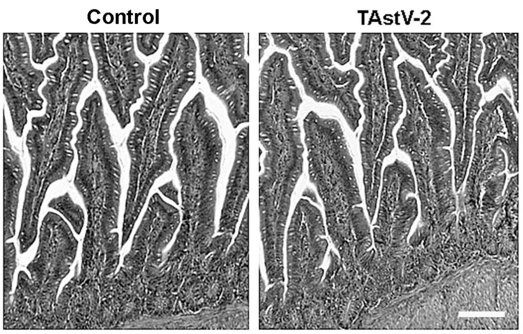
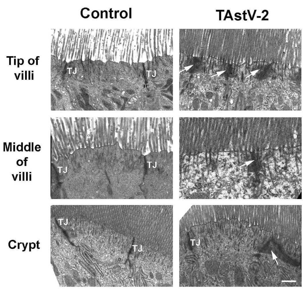
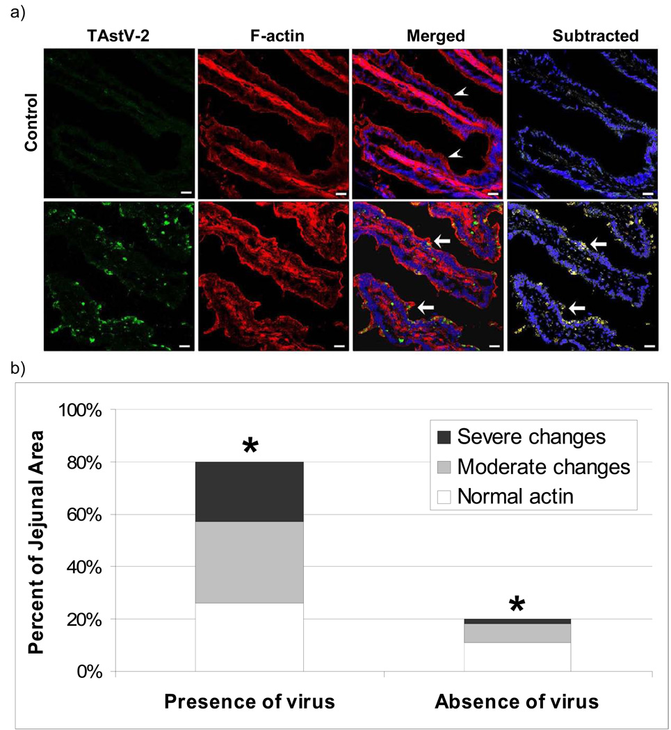
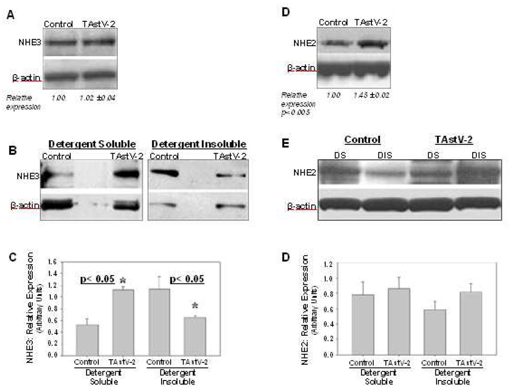
Astroviruses are known to be a leading cause of diarrhea in infants and the immunocompromised; however, our understanding of this endemic pathogen is limited. Histological analyses of astrovirus pathogenesis demonstrate clinical disease is not associated with changes to intestinal architecture, inflammation, or cell death. Recent studies in vitro have suggested that astroviruses induce actin rearrangement leading to loss of barrier function. The current study used the type-2 turkey astrovirus (TAstV-2) and turkey poult model of astrovirus disease to examine how astrovirus infection affects the ultrastructure and electrophysiology of the intestinal epithelium. These data demonstrate that infection results in changes to the epithelial ultrastructure, rearrangement of F-actin, decreased absorption of sodium, as well as redistribution of the sodium/hydrogen exchanger 3 (NHE3) from the membrane to the cytoplasm. Collectively, these data suggest astrovirus infection induces sodium malabsorption, possibly through redistribution of specific sodium transporters, which results in the development of an osmotic diarrhea.
2010 Elsevier Inc. All rights reserved.
Figures
Similar articles
-
Oral Administration of Astrovirus Capsid Protein Is Sufficient To Induce Acute Diarrhea In Vivo.mBio. 2016 Nov 1;7(6):e01494-16. doi: 10.1128/mBio.01494-16. mBio. 2016. PMID: 27803180 Free PMC article.
-
The role of type-2 turkey astrovirus in poult enteritis syndrome.Poult Sci. 2011 Dec;90(12):2747-52. doi: 10.3382/ps.2011-01617. Poult Sci. 2011. PMID: 22080013 Free PMC article.
-
Astrovirus induces diarrhea in the absence of inflammation and cell death.J Virol. 2003 Nov;77(21):11798-808. doi: 10.1128/jvi.77.21.11798-11808.2003. J Virol. 2003. PMID: 14557664 Free PMC article.
-
Pathogenesis of astrovirus infection.Viral Immunol. 2005;18(1):4-10. doi: 10.1089/vim.2005.18.4. Viral Immunol. 2005. PMID: 15802949 Review.
-
A Review of the Emerging Poultry Visceral Gout Disease Linked to Avian Astrovirus Infection.Int J Mol Sci. 2022 Sep 9;23(18):10429. doi: 10.3390/ijms231810429. Int J Mol Sci. 2022. PMID: 36142340 Free PMC article. Review.
Cited by
-
Characterizing a Murine Model for Astrovirus Using Viral Isolates from Persistently Infected Immunocompromised Mice.J Virol. 2019 Jun 14;93(13):e00223-19. doi: 10.1128/JVI.00223-19. Print 2019 Jul 1. J Virol. 2019. PMID: 30971471 Free PMC article.
-
Human rotavirus strain Wa downregulates NHE1 and NHE6 expressions in rotavirus-infected Caco-2 cells.Virus Genes. 2017 Jun;53(3):367-376. doi: 10.1007/s11262-017-1444-0. Epub 2017 Mar 13. Virus Genes. 2017. PMID: 28289928
-
Oral Administration of Astrovirus Capsid Protein Is Sufficient To Induce Acute Diarrhea In Vivo.mBio. 2016 Nov 1;7(6):e01494-16. doi: 10.1128/mBio.01494-16. mBio. 2016. PMID: 27803180 Free PMC article.
-
Molecular Characterization and Determination of Relative Cytokine Expression in Naturally Infected Day-Old Chicks with Chicken Astrovirus Associated to White Chick Syndrome.Animals (Basel). 2020 Jul 14;10(7):1195. doi: 10.3390/ani10071195. Animals (Basel). 2020. PMID: 32674433 Free PMC article.
-
Treatment of diarrhoea in rural African communities: an overview of measures to maximise the medicinal potentials of indigenous plants.Int J Environ Res Public Health. 2012 Oct 26;9(11):3911-33. doi: 10.3390/ijerph9113911. Int J Environ Res Public Health. 2012. PMID: 23202823 Free PMC article. Review.
References
-
- Akhter S, Kovbasnjuk O, Li X, Cavet M, Noel J, Arpin M, Hubbard AL, Donowitz M. Na(+)/H(+) exchanger 3 is in large complexes in the center of the apical surface of proximal tubule-derived OK cells. Am J Physiol Cell Physiol. 2002;283:C927–C940. - PubMed
-
- Argenzio RA, Armstrong M. ANP inhibits NaCl absorption and elicits Cl secretion in porcine colon: evidence for cGMP and Ca mediation. Am J Physiol. 1993;265:R57–R65. - PubMed
-
- Argenzio RA, Liacos JA. Endogenous prostanoids control ion transport across neonatal porcine ileum in vitro. Am J Vet Res. 1990;51:747–751. - PubMed
-
- Bachmann O, Riederer B, Rossmann H, Groos S, Schultheis PJ, Shull GE, Gregor M, Manns MP, Seidler U. The Na+/H+ exchanger isoform 2 is the predominant NHE isoform in murine colonic crypts and its lack causes NHE3 upregulation. Am J Physiol Gastrointest Liver Physiol. 2004;287:G125–G133. - PubMed
-
- Behling-Kelly E, Schultz-Cherry S, Koci M, Kelley L, Larsen D, Brown C. Localization of astrovirus in experimentally infected turkeys as determined by in situ hybridization. Vet Pathol. 2002;39:595–598. - PubMed
Publication types
MeSH terms
Substances
Grants and funding
LinkOut - more resources
Full Text Sources
