

A comparison of the influence of material on in vitro cartilage tissue engineering with PCL, PGS, and POC 3D scaffold architecture seeded with chondrocytes
- PMID: 20219243
- PMCID: PMC4367812
- DOI: 10.1016/j.biomaterials.2010.01.145
A comparison of the influence of material on in vitro cartilage tissue engineering with PCL, PGS, and POC 3D scaffold architecture seeded with chondrocytes
Abstract
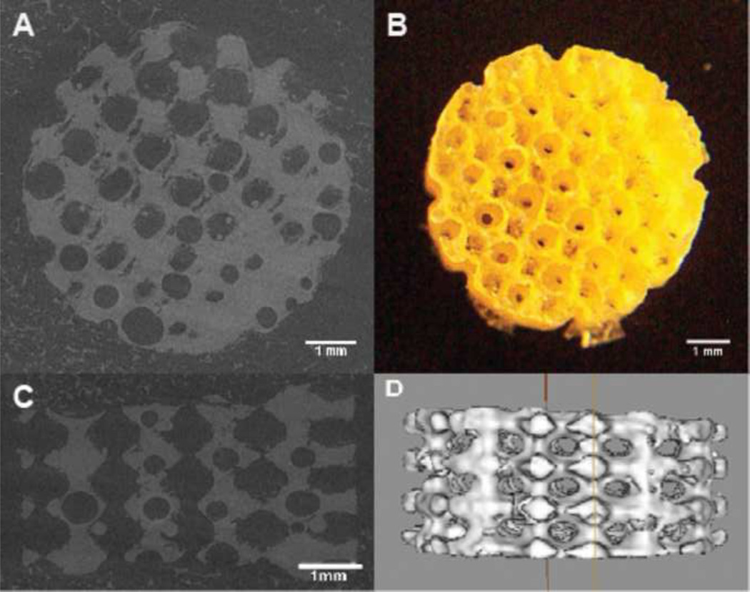
The goal of this study was to determine material effects on cartilage regeneration for scaffolds with the same controlled architecture. The 3D polycaprolactone (PCL), poly (glycerol sebacate) (PGS), and poly (1,8 octanediol-co-citrate) (POC) scaffolds of the same design were physically characterized and tissue regeneration in terms of cell phenotype, cellular proliferation and differentiation, and matrix production were compared to find which material would be most optimal for cartilage regeneration in vitro. POC provided the best support for cartilage regeneration in terms of tissue ingrowth, matrix production, and relative mRNA expressions for chondrocyte differentiation (Col2/Col1). PGS was seen as the least favorable material for cartilage based on its relatively high de-differentiation (Col1), hypertrophic mRNA expression (Col10) and high matrix degradation (MMP13, MMP3) results. PCL still provided microenvironments suitable for cells to be active yet it seemed to cause de-differentiation (Col1) of chondrocytes inside the scaffold while many cells migrated out, growing cartilage outside the scaffold.
Copyright (c) 2010 Elsevier Ltd. All rights reserved.
Figures


Similar articles
-
Biomimetic poly(glycerol sebacate)/polycaprolactone blend scaffolds for cartilage tissue engineering.J Mater Sci Mater Med. 2019 Apr 29;30(5):53. doi: 10.1007/s10856-019-6257-3. J Mater Sci Mater Med. 2019. PMID: 31037512
-
A new biodegradable polyester elastomer for cartilage tissue engineering.J Biomed Mater Res A. 2006 May;77(2):331-9. doi: 10.1002/jbm.a.30607. J Biomed Mater Res A. 2006. PMID: 16404714
-
Effect of biodegradation and de novo matrix synthesis on the mechanical properties of valvular interstitial cell-seeded polyglycerol sebacate-polycaprolactone scaffolds.Acta Biomater. 2013 Apr;9(4):5963-73. doi: 10.1016/j.actbio.2012.11.014. Epub 2012 Nov 17. Acta Biomater. 2013. PMID: 23168222 Free PMC article.
-
Hybrid and Composite Scaffolds Based on Extracellular Matrices for Cartilage Tissue Engineering.Tissue Eng Part B Rev. 2019 Jun;25(3):202-224. doi: 10.1089/ten.TEB.2018.0245. Tissue Eng Part B Rev. 2019. PMID: 30648478 Review.
-
Cartilage tissue engineering.Adv Exp Med Biol. 2004;553:317-29. doi: 10.1007/978-0-306-48584-8_24. Adv Exp Med Biol. 2004. PMID: 15503466 Review. No abstract available.
Cited by
-
Polyglycerol Hyperbranched Polyesters: Synthesis, Properties and Pharmaceutical and Biomedical Applications.Int J Mol Sci. 2019 Dec 9;20(24):6210. doi: 10.3390/ijms20246210. Int J Mol Sci. 2019. PMID: 31835372 Free PMC article. Review.
-
Harnessing cell–biomaterial interactions for osteochondral tissue regeneration.Adv Biochem Eng Biotechnol. 2012;126:67-104. doi: 10.1007/10_2011_107. Adv Biochem Eng Biotechnol. 2012. PMID: 21975954 Free PMC article.
-
Synthesis and Properties of Magnetic Fe3O4/PCL Porous Biocomposite Scaffolds with Different Sizes and Quantities of Fe3O4 Particles.Bioengineering (Basel). 2022 Jun 26;9(7):278. doi: 10.3390/bioengineering9070278. Bioengineering (Basel). 2022. PMID: 35877329 Free PMC article.
-
Photopolymerizable Biomaterials and Light-Based 3D Printing Strategies for Biomedical Applications.Chem Rev. 2020 Oct 14;120(19):10695-10743. doi: 10.1021/acs.chemrev.9b00810. Epub 2020 Apr 23. Chem Rev. 2020. PMID: 32323975 Free PMC article. Review.
-
Strategic design and fabrication of engineered scaffolds for articular cartilage repair.J Funct Biomater. 2012 Nov 14;3(4):799-838. doi: 10.3390/jfb3040799. J Funct Biomater. 2012. PMID: 24955748 Free PMC article.
References
-
- Kemppainen JM, Hollister SJ. Differential effects of designed scaffold permeability on chondrogenesis by chondrocytes and bone marrow stromal cells. Biomaterials. 2010 Jan;31(2):279–287. Epub 2009 Oct 8. - PubMed
-
- Kim HJ, Lee JH, Im GI. Chondrogenesis using mesenchymal stem cells and PCL scaffolds. J Biomed Mater Res A. 2010 Feb;92(2):659–666. - PubMed
-
- Izquierdo R, Garcia-Giralt N, Rodriguez MT, Caceres E, Garcia SJ, Gomez Ribelles JL, Monleon M, Monllau JC, Suay J. Biodegradable PCL scaffolds with an interconnected spherical pore network for tissue engineering. J Biomed Mater Res A. 2008;85:25–35. - PubMed
-
- Li WJ, Tuli R, Okafor C, Derfoul A, Danielson KG, Hall DJ, Tuan RS. A three-dimensional nanofibrous scaffold for cartilage tissue engineering using human mesenchymal stem cells. Biomaterials. 2005;26:599–609. - PubMed
-
- Kemppainen JM, Hollister SJ. Tailoring the mechanical properties of 3D-designed poly(glycerol sebacate) scaffolds for cartilage applications. J Biomed Mater Res A. 2010 Jan 20; - PubMed
Publication types
MeSH terms
Substances
Grants and funding
LinkOut - more resources
Full Text Sources
Miscellaneous
