

Establishing a model spinal cord injury in the African green monkey for the preclinical evaluation of biodegradable polymer scaffolds seeded with human neural stem cells
- PMID: 20219534
- PMCID: PMC4157751
- DOI: 10.1016/j.jneumeth.2010.02.019
Establishing a model spinal cord injury in the African green monkey for the preclinical evaluation of biodegradable polymer scaffolds seeded with human neural stem cells
Abstract
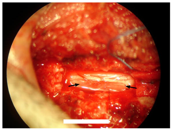
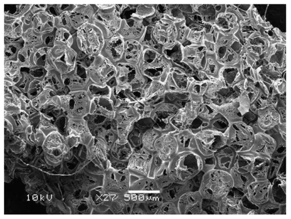
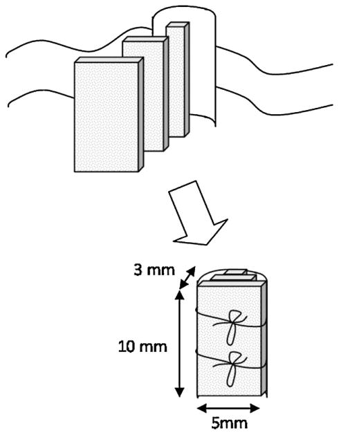
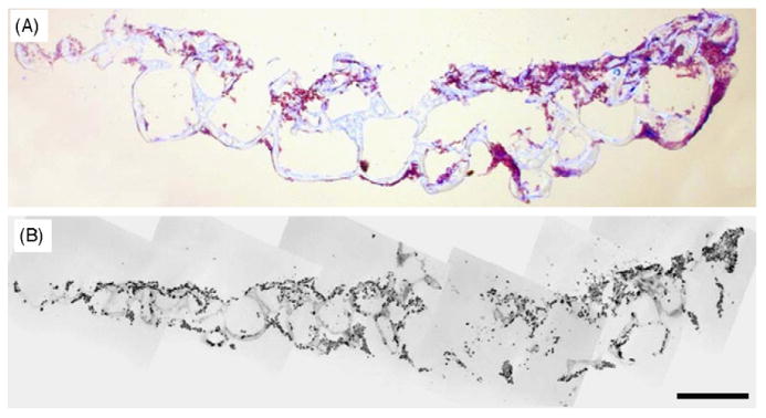
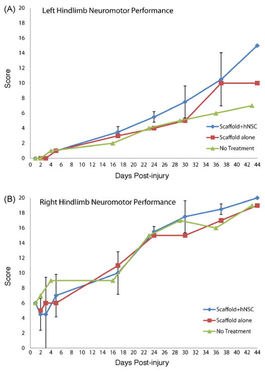
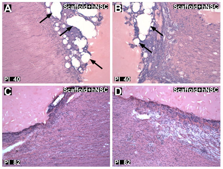
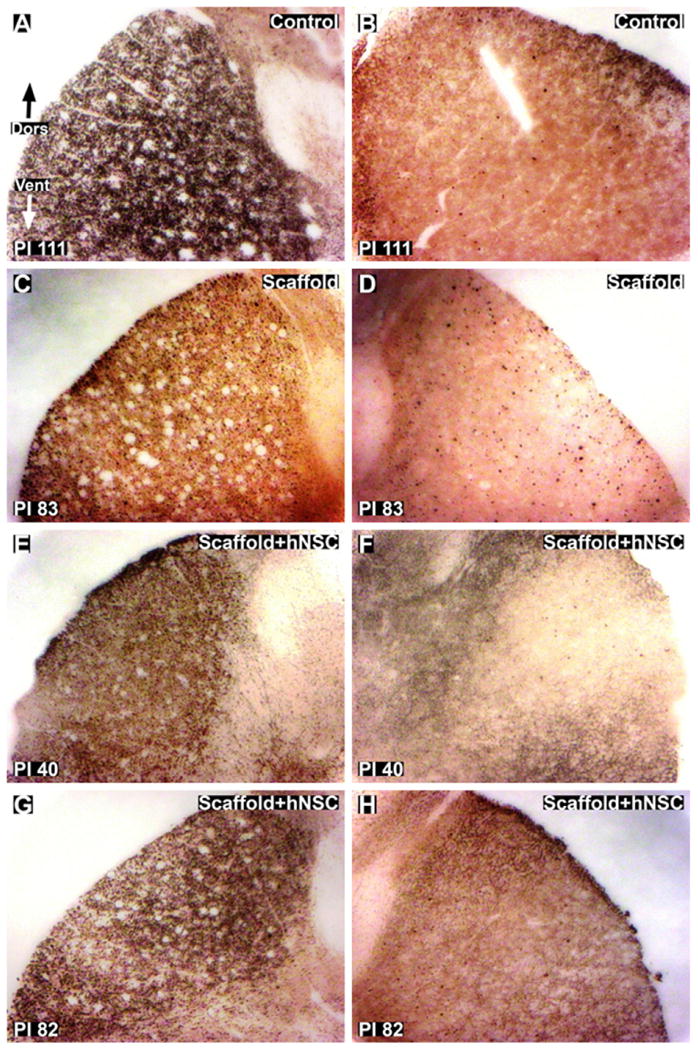
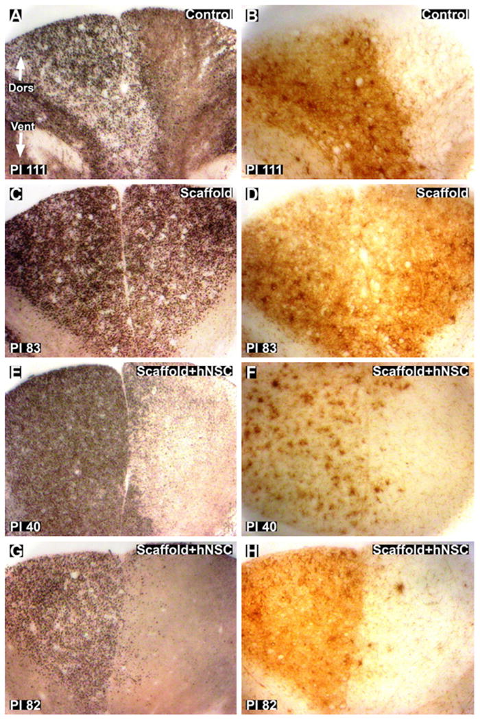
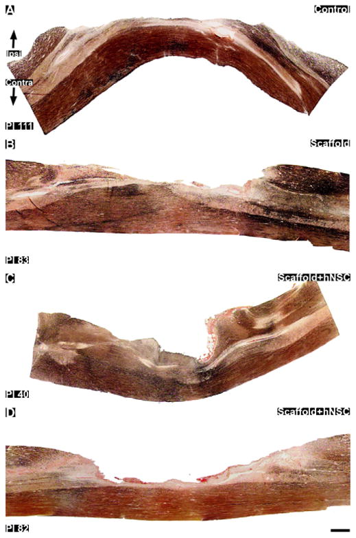
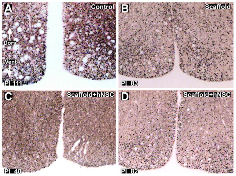
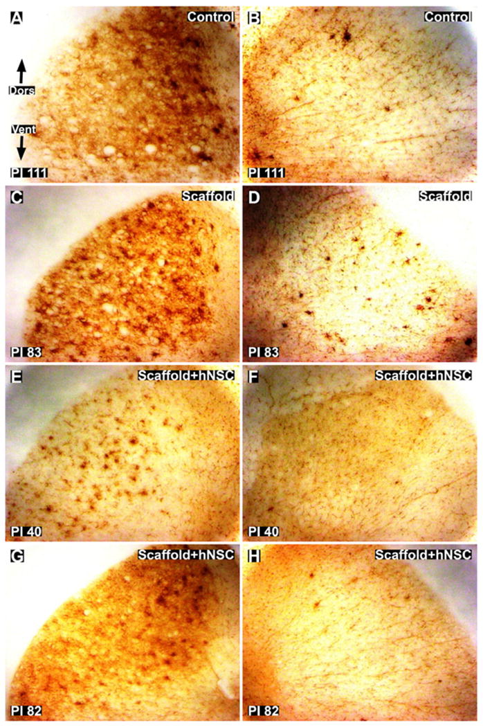
Given the involvement of post-mitotic neurons, long axonal tracts and incompletely elucidated injury and repair pathways, spinal cord injury (SCI) presents a particular challenge for the creation of preclinical models to robustly evaluate longitudinal changes in neuromotor function in the setting in the presence and absence of intervention. While rodent models exhibit high degrees of spontaneous recovery from SCI injury, animal care concerns preclude complete cord transections in non-human primates and other larger vertebrate models. To overcome such limitations a segmental thoracic (T9-T10) spinal cord hemisection was created and characterized in the African green monkey. Physiological tolerance of the model permitted behavioral analyses for a prolonged period post-injury, extending to predefined study termination points at which histological and immunohistochemical analyses were performed. Four monkeys were evaluated (one receiving no implant at the lesion site, one receiving a poly(lactide-co-glycolide) (PLGA) scaffold, and two receiving PLGA scaffolds seeded with human neural stem cells (hNSC)). All subjects exhibited Brown-Séquard syndrome 2 days post-injury consisting of ipsilateral hindlimb paralysis and contralateral hindlimb hypesthesia with preservation of bowel and bladder function. A 20-point observational behavioral scoring system allowed quantitative characterization of the levels of functional recovery. Histological endpoints including silver degenerative staining and Iba1 immunohistochemistry, for microglial and macrophage activation, were determined to reliably define lesion extent and correlate with neurobehavioral data, and justify invasive telemetered electromyographic and kinematic studies to more definitively address efficacy and mechanism.
Copyright (c) 2010 Elsevier B.V. All rights reserved.
Figures
Similar articles
-
Transplantation of galectin-1-expressing human neural stem cells into the injured spinal cord of adult common marmosets.J Neurosci Res. 2010 May 15;88(7):1394-405. doi: 10.1002/jnr.22322. J Neurosci Res. 2010. PMID: 20091712
-
Effects of human neural stem cell transplantation in canine spinal cord hemisection.Neurol Res. 2009 Nov;31(9):996-1002. doi: 10.1179/174313209X385626. Epub 2009 Jan 9. Neurol Res. 2009. PMID: 19138477
-
Biodegradable scaffolds promote tissue remodeling and functional improvement in non-human primates with acute spinal cord injury.Biomaterials. 2017 Apr;123:63-76. doi: 10.1016/j.biomaterials.2017.01.024. Epub 2017 Jan 25. Biomaterials. 2017. PMID: 28167393
-
Neuronal repair and replacement in spinal cord injury.J Neurol Sci. 2008 Feb 15;265(1-2):63-72. doi: 10.1016/j.jns.2007.05.004. Epub 2007 Jun 12. J Neurol Sci. 2008. PMID: 17568612 Review.
-
Cell-based transplantation strategies to promote plasticity following spinal cord injury.Exp Neurol. 2012 May;235(1):78-90. doi: 10.1016/j.expneurol.2011.02.010. Epub 2011 Feb 17. Exp Neurol. 2012. PMID: 21333647 Review.
Cited by
-
Advances in Neural Stem Cell Therapy for Spinal Cord Injury: Safety, Efficacy, and Future Perspectives.Neurospine. 2022 Dec;19(4):946-960. doi: 10.14245/ns.2244658.329. Epub 2022 Nov 10. Neurospine. 2022. PMID: 36351442 Free PMC article.
-
Clinical and experimental advances in regeneration of spinal cord injury.J Tissue Eng. 2010 Nov 2;2010:650857. doi: 10.4061/2010/650857. J Tissue Eng. 2010. PMID: 21350645 Free PMC article.
-
Transplanting neural progenitor cells to restore connectivity after spinal cord injury.Nat Rev Neurosci. 2020 Jul;21(7):366-383. doi: 10.1038/s41583-020-0314-2. Epub 2020 Jun 9. Nat Rev Neurosci. 2020. PMID: 32518349 Free PMC article. Review.
-
Animal Models of Spinal Cord Injury.Biomedicines. 2025 Jun 10;13(6):1427. doi: 10.3390/biomedicines13061427. Biomedicines. 2025. PMID: 40564146 Free PMC article. Review.
-
Methods for functional assessment after C7 spinal cord hemisection in the rhesus monkey.Neurorehabil Neural Repair. 2012 Jul-Aug;26(6):556-69. doi: 10.1177/1545968311421934. Epub 2012 Feb 13. Neurorehabil Neural Repair. 2012. PMID: 22331214 Free PMC article.
References
-
- Babu RS, Muthusamy R, Namasivayam A. Behavioural assessment of functional recovery after spinal cord hemisection in the bonnet monkey. Journal of Neurological Sciences. 2000;178:136–52. - PubMed
-
- Baptiste DC, Fehlings MG. Update on the treatment of spinal cord injury. Progress in Brain Research. 2007;161:217–33. - PubMed
-
- Basso DM, Beattie MS, Bresnahan JC. Graded histological and locomotor outcomes after spinal cord contusion using the NYU weight-drop device versus transaction. Experimental Neurology. 1996;139:244–56. - PubMed
-
- Brown-Séquard CÉ. De la transmission croisée des impressions sensitives par la moelle épinière. Comptes rendus de la Société de biologie. 1851;2:33–44.
-
- Courtine G, Roy RR, Hodgson J, McKay H, Raven J, Zhong H, Yang H, Tuszynski MH, Edgerton VR. Kinematic and EMG determinants in quadrupedal locomotion of non-human primate (rhesus) Journal of Neurophysiology. 2005;93:3127–45. - PubMed
Publication types
MeSH terms
Grants and funding
LinkOut - more resources
Full Text Sources
Medical
Research Materials
