. 2010 Jun;61(6):1683-97.
doi: 10.1093/jxb/erq039. Epub 2010 Mar 10.

Removing the mustard oil bomb from seeds: transgenic ablation of myrosin cells in oilseed rape (Brassica napus) produces MINELESS seeds

Affiliations

Removing the mustard oil bomb from seeds: transgenic ablation of myrosin cells in oilseed rape (Brassica napus) produces MINELESS seeds

Birgit Hafeld Borgen et al. J Exp Bot. 2010 Jun.

Abstract

Many plant phytochemicals constitute binary enzyme-glucoside systems and function in plant defence. In brassicas, the enzyme myrosinase is confined to specific myrosin cells that separate the enzyme from its substrate; the glucosinolates. The myrosinase-catalysed release of toxic and bioactive compounds such as isothiocyanates, upon activation or tissue damage, has been termed 'the mustard oil bomb' and characterized as a 'toxic mine' in plant defence. The removal of myrosin cells and the enzyme that triggers the release of phytochemicals have been investigated by genetically modifying Brassica napus plants to remove myrosinase-storing idioblasts. A construct with the seed myrosin cell-specific Myr1.Bn1 promoter was used to express a ribonuclease, barnase. Transgenic plants ectopically expressing barnase were embryo lethal. Co-expressing barnase under the control of the Myr1.Bn1 promoter with the barnase inhibitor, barstar, under the control of the cauliflower mosaic virus 35S promoter enabled a selective and controlled death of myrosin cells without affecting plant viability. Ablation of myrosin cells was confirmed with light and electron microscopy, with immunohistological analysis and immunogold-electron microscopy analysis showing empty holes where myrosin cells normally are localized. Further evidence for a successful myrosin cell ablation comes from immunoblots showing absence of myrosinase and negligible myrosinase activity, and autolysis experiments showing negligible production of glucosinolate hydrolysis products. The plants where the myrosin defence cells have been ablated and named 'MINELESS plants'. The epithiospecifier protein profile and glucosinolate levels were changed in MINELESS plants, pointing to localization of myrosinases and a 35 kDa epithiospecifier protein in myrosin cells and a reduced turnover of glucosinolates in MINELESS plants.


Figures

Fig. 1.
Fig. 1.
Map of the myrosinase promoter (pMyr1 as a HindIII–NcoI 2923 bp fragment) transformation constructs in pBI101. (A) Myr1.Bn1 promoter:Barnase fusion (Barnase:3′NOS as a NcoI–EcoRI fragment). (B) Myr1.Bn1 promoter:Barnase–35S:Barstar (35S:Barstar EcoRI–EcoRI as an orientation-selected insert) double fusion used to ablate myrosin cells in Brassica napus seeds. LB, left border; RB, right border, 3′NOS, nopaline synthase terminator; NPTII, kanamycin selection; 3′g7, g7 terminator; BARN, barnase gene; BAR*, barstar gene; 35S, CaMV promoter, restriction sites, and total size (bp) of the constructs. Arrows denote transcriptional orientation.
Fig. 2.
Fig. 2.
Structural analysis of myrosin cells from semi-thin sections of wild type and MINELESS seeds by light microscopy. (A and B) Sections from MINELESS seeds stained with toluidine blue where the insert in (B) shows a normal myrosin cell from the wild type marked with a star. (C–E) Sections labelled with anti-myrosinase polyclonal antibody (K089) and FITC-conjugated secondary antibodies. (C) Wild type section; (D and E) MINELESS sections. White arrows indicate ablated or normal myrosin cells. Bars=10 μm. The exposure time in D and E was increased to visualize the ablated myrosin cells.
Fig. 3.
Fig. 3.
Transmission electron microscopic structural analysis of myrosin cells from thin sections of wild type and MINELESS seeds. (A) Unlabelled section from wild type seeds showing a myrosin cell with the typical granulated matrix inside myrosin grains marked by stars. (B–E) Immunogold-EM sections labelled with anti-myrosinase (K089) and 15 nm colloidal gold. (B–C, E) MINELESS sections showing different stages of myrosin cell ablation. (D) Wild type section. Myrosin grains in the wild type (D) are marked by stars and ablated myrosin cells in MINELESS (C, E) are marked by starbursts. Inserts demonstrate specific detection of myrosinase inside a myrosin grain (D) and specific ablation inside a myrosin cell (E). Bars=2 μm.
Fig. 4.
Fig. 4.
Distribution of specific myrosinase activity in wild type and MINELESS single seeds.
Fig. 5.
Fig. 5.
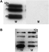
Immunoblot analysis of wild type (W) and MINELESS (M) single seeds. (A) Expression of myrosinase proteins was detected with anti-myrosinase 3D7 antibodies. (B) Expression of myrosinase-binding proteins (MBPs) was detected with 34:14 antibodies. A 10 μg aliquot of total protein was loaded in each lane.
Fig. 6.
Fig. 6.
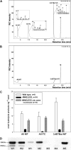
Epithiospecifier protein (ESP) activity assay by assessing sinigrin hydrolysis product formation and immunoblot analysis of ESP expression from single wild type and MINELESS seeds. (A) GC chromatogram of a wild type single seed showing sinigrin hydrolysis products. (B) GC chromatogram of a MINELESS single seed showing sinigrin hydrolysis products. (C) Total hydrolysis products produced in a wild type single seed, a MINELESS single seed, and a single MINELESS seed supplemented with purified myrosinase. Error bars represent SE. (D) ESP expression in wild type (W1 and W2) and MINELESS (M1–M5) single seeds. A 10 μg aliquot of total protein was loaded in each lane. The expression of ESP was detected with the anti-ESP antibody as described by Foo et al. (2000).
Fig. 7.
Fig. 7.
Myrosinase activity and glucosinolates from half a seed of the wild type and MINELESS. (A) Myrosinase activity. (B) Aliphatic, indolic, and total glucosinolate content. (C) Glucosinolate profile and content of individual glucosinolates. Error bars represent the SE.
Fig. 8.
Fig. 8.
Scatter plot showing glucosinolate content versus myrosinase activity from wild type and MINELESS single seeds.

Similar articles

Cited by

References

    1. Ahuja I, Rohloff J, Bones AM. Defence mechanisms of Brassicaceae: implications for plant–insect interactions and potential for integrated pest management. A review. Agronomy for Sustainable Development. 2009 DOI: 10.1051/agro/2009025.
    1. Andreasson E, Jorgensen LB, Hoglund AS, Rask L, Meijer J. Different myrosinase and idioblast distribution in Arabidopsis and Brassica napus. Plant Physiology. 2001;127:1750–1763. - PMC - PubMed
    1. Barth C, Jander G. Arabidopsis myrosinases TGG1 and TGG2 have redundant function in glucosinolate breakdown and insect defense. The Plant Journal. 2006;46:549–562. - PubMed
    1. Beals TP, Goldberg RB. A novel cell ablation strategy blocks tobacco anther dehiscence. The Plant Cell. 1997;9:1527–1545. - PMC - PubMed
    1. Bednarek P, Pislewska-Bednarek M, et al. A glucosinolate metabolism pathway in living plant cells mediates broad-spectrum antifungal defense. Science. 2009;323:101–106. - PubMed

Publication types

MeSH terms