Review
. 2010 May;298(5):R1173-87.
doi: 10.1152/ajpregu.00735.2009. Epub 2010 Mar 10.

Regulatory interactions between muscle and the immune system during muscle regeneration

Affiliations
Review

Regulatory interactions between muscle and the immune system during muscle regeneration

James G Tidball et al. Am J Physiol Regul Integr Comp Physiol. 2010 May.

Abstract

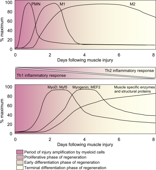
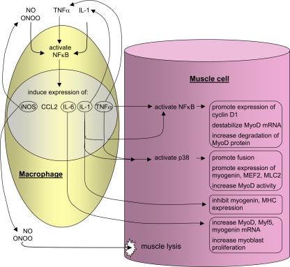
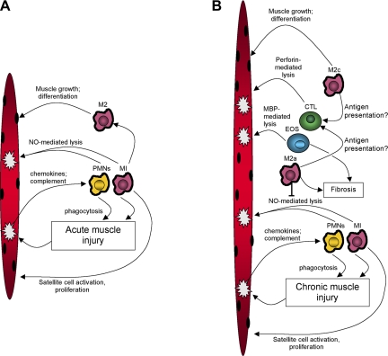
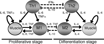
Recent discoveries reveal complex interactions between skeletal muscle and the immune system that regulate muscle regeneration. In this review, we evaluate evidence that indicates that the response of myeloid cells to muscle injury promotes muscle regeneration and growth. Acute perturbations of muscle activate a sequence of interactions between muscle and inflammatory cells. The initial inflammatory response is a characteristic Th1 inflammatory response, first dominated by neutrophils and subsequently by CD68(+) M1 macrophages. M1 macrophages can propagate the Th1 response by releasing proinflammatory cytokines and cause further tissue damage through the release of nitric oxide. Myeloid cells in the early Th1 response stimulate the proliferative phase of myogenesis through mechanisms mediated by TNF-alpha and IL-6; experimental prolongation of their presence is associated with delayed transition to the early differentiation stage of myogenesis. Subsequent invasion by CD163(+)/CD206(+) M2 macrophages attenuates M1 populations through the release of anti-inflammatory cytokines, including IL-10. M2 macrophages play a major role in promoting growth and regeneration; their absence greatly slows muscle growth following injury or modified use and inhibits muscle differentiation and regeneration. Chronic muscle injury leads to profiles of macrophage invasion and function that differ from acute injuries. For example, mdx muscular dystrophy yields invasion of muscle by M1 macrophages, but their early invasion is accompanied by a subpopulation of M2a macrophages. M2a macrophages are IL-4 receptor(+)/CD206(+) cells that reduce cytotoxicity of M1 macrophages. Subsequent invasion of dystrophic muscle by M2c macrophages is associated with progression of the regenerative phase in pathophysiology. Together, these findings show that transitions in macrophage phenotype are an essential component of muscle regeneration in vivo following acute or chronic muscle damage.


Figures

Fig. 1.
Fig. 1.
Inflammatory lesion in skeletal muscle shows codistribution of M1 and M2 macrophages with activated satellite cells. Anti-F4/80 (red fluorophore) binds all macrophages. Anti-CD206 (green fluorophore) binds M2 macrophages and satellite cells. Hoechts labeling (blue) shows nuclei. CD206−/F4/80+ cells (red) are M1 macrophages. CD206+/F4/80+ cells (orange) are M2 macrophages. CD206+/F4/80− cells (green) are satellite cells. Muscle section was obtained from 4-wk-old mdx quadriceps muscle. Scale bar = 50 μm.
Fig. 2.
Fig. 2.
Generalized time course of changes in myeloid cell populations (top) and changes in the expression levels of muscle-specific transcription factors, enzymes, and structural proteins in muscle (bottom) following acute muscle injury. The transition from a Th1 inflammatory response to a Th2 inflammatory response coincides with a transition from the early proliferative stage of myogenesis to the early and terminal stages of myogenesis. Experimental perturbations that disrupt the Th1 to Th2 transition also disrupt the transition from proliferative to differentiation stages of myogenesis, suggesting a functional linkage between differentiation in the myeloid compartment and myogenic compartment. PMN, neutrophils; M1, M1 macrophages; M2, M2 macrophages.
Fig. 3.
Fig. 3.
Activation of NF-κB in muscle cells or macrophages can directly or indirectly influence muscle cell proliferation and differentiation. Th1 cytokines or oxidative stress can increase NF-κB activation in muscle or macrophages. Those cytokines can then contribute to further activation of NF-κB or they can act on muscle cells to affect their proliferation or differentiation. In general, NF-κB activation increases proliferation and inhibits differentiation, increasing the expression of transcripts needed for cell cycle progression and by decreasing the expression or destabilizing transcripts needed for muscle to experience terminal differentiation. NO, nitric oxide; iNOS, inducible nitric oxide synthesis; ONOO, peroxynitrate; MEF2, myocyte enhancer binding factor-2; MHC, myson heavy chain.
Fig. 4.
Fig. 4.
Summary of general interactions between immune cells and muscle cells following acute muscle injury (A) or during chronic muscle injury that occurs in mdx dystrophy (B). A: acute damage causes release of chemoattractant molecules that initially attract neutrophils (PMNs) or M1 macrophages (M1) into the muscle. Neutrophils and M1 macrophages promote further damage through nitric oxide (NO)-mediated processes. M1 macrophages also release cytokines that can promote satellite cell activation and proliferation. Neutrophils and M1 macrophages are then replaced by M2 macrophages that then promote muscle repair, differentiation and growth. B: during chronic muscle injury, the initial inflammatory response is similar to the response to acute damage, typified by neutrophil and M1 macrophage invasion. However, M2a macrophages invade contemporaneously and inhibit NO-mediated lysis by M1 macrophages and promote muscle fibrosis through arginase metabolism of arginine. M2 macrophages may also participate in activation of cytotoxic T-cells that are then able to promote muscle damage through perforin-mediated processes. The Th2 inflammatory environment also increases the activation of eosinophils (EOS) that contribute to muscle lysis through major basic protein-1 (MBP-1) mediated lysis. Eosinophils also increase muscle fibrosis through MBP-dependent processes and negatively regulate the cellular immune response to chronically injured muscle.
Fig. 5.
Fig. 5.
Generalized diagram of the interactions between macrophages and muscle cells during the proliferative stage and differentiation stages of myogenesis in injured muscle. “Th1” and “Th2” represent cells that produce Th1 or Th2 cytokines, that include helper T-cells or macrophages. “M1” and “M2” represent macrophage populations. Solid lines indicate activating effects on target cells. Dotted lines represent deactivating effects.

Similar articles

Cited by

References

    1. Abood EA, Jones MM. Macrophages in developing mammalian skeletal muscle: evidence for muscle fibre death as a normal developmental event. Acta Anat (Basel) 140: 201–212, 1991 - PubMed
    1. Arnold L, Henry A, Poron F, Baba-Amer Y, van Rooijen N, Plonquet A, Gherardi RK, Chazaud B. Inflammatory monocytes recruited after skeletal muscle injury switch into anti-inflammatory macrophages to support myogenesis. J Exp Med 204: 1057–1069, 2007 - PMC - PubMed
    1. Backman E, Henriksson KG. Low-dose prednisolone treatment in Duchenne and Becker muscular dystrophy. Neuromusc Disord 5: 233–241, 1995 - PubMed
    1. Bakkar N, Wang J, Ladner KJ, Wang H, Dahlman JM, Carathers M, Acharyya S, Rudnicki MA, Hollenbach AD, Guttridge DC. IKK/NF-κB regulates skeletal myogenesis via a signaling switch to inhibit differentiation and promote mitochondrial biogenesis. J Cell Biol 180: 787–802, 2008 - PMC - PubMed
    1. Bartoli C, Civatte M, Pellissier JF, Figarella-Branger D. CCR2A and CCR2B, the two isoforms of the monocyte chemoattractant protein-1 receptor are up-regulated and expressed by different cell subsets in idiopathic inflammatory myopathies. Acta Neuropathol (Berl) 102: 385–392, 2001 - PubMed

Publication types