Review
. 2010 Jun;298(6):R1449-67.
doi: 10.1152/ajpregu.00130.2010. Epub 2010 Mar 10.

The Walter B. Cannon Memorial Award Lecture, 2009. Physiology in perspective: The wisdom of the body. In search of autonomic balance: the good, the bad, and the ugly

Affiliations
Review

The Walter B. Cannon Memorial Award Lecture, 2009. Physiology in perspective: The wisdom of the body. In search of autonomic balance: the good, the bad, and the ugly

François M Abboud. Am J Physiol Regul Integr Comp Physiol. 2010 Jun.

Abstract

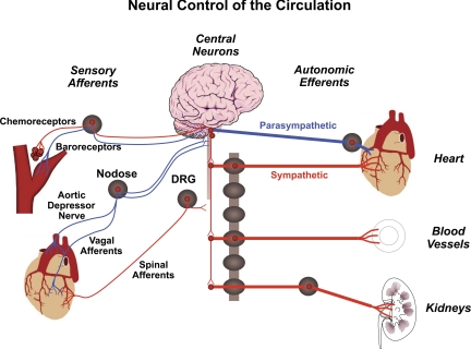
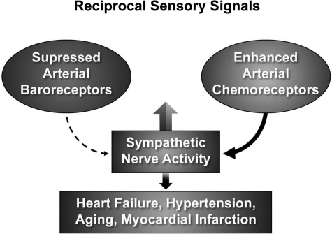
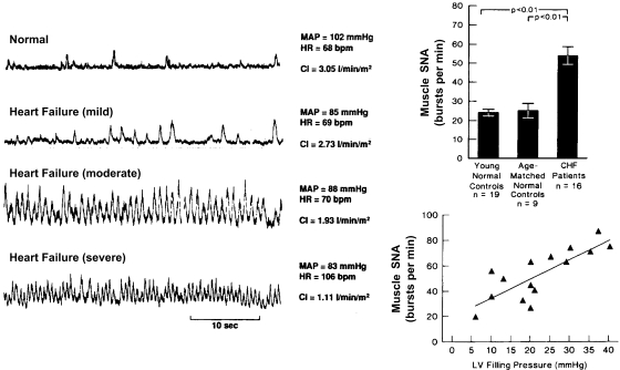
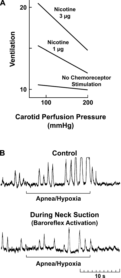
Walter B. Cannon's research on the sympathetic nervous system and neurochemical transmission was pioneering. Wisdom has endowed our body with a powerful autonomic neural regulation of the circulation that provides optimal perfusion of every organ in accordance to its metabolic needs. Exquisite sensors tuned to an optimal internal environment trigger central and peripheral sympathetic and parasympathetic motor neurons and allow desirable and beneficial adjustments to physiologic needs as well as to acute cardiovascular stresses. This short review, presented as The Walter B. Cannon Memorial Award Lecture for 2009, addresses the mechanisms that disrupt sensory signaling and result in a chronic maladjustment of the autonomic neural output that in many cardiovascular diseases results in excessive increases in the risks of dying. The hopes for any reduction of those risks resides in an understanding of the molecular determinants of neuronal signaling.


Figures

Fig. 1.
Fig. 1.
Neural control of the circulation. Two major sensory signals are: 1) baroreceptors and mechanosensory sympathoinhibition, and 2) chemoreceptors and chemosensory sympathoexcitation. DRG, dorsal root ganglion.
Fig. 2.
Fig. 2.
The augmented sympathetic nerve activity (SNA) in hypertension, heart failure, myocardial infarction, diabetes, and aging may result in large part from impaired baroreceptor activity (dashed-lined arrow) and enhanced chemoreceptor activity (solid-line arrow). Both increase SNA.
Fig. 3.
Fig. 3.
Direct recordings of muscle SNA in patients with congestive heart failure (CHF) show dramatic increases in severe failure. The increases in SNA correlate with increases in left ventricular (LV) filling pressures and are significantly greater than in young and age-matched normal controls (bar graph). MAP, mean arterial pressure; HR, heart rate. [From Ferguson et al. (21) and Leimbach et al. (43).]
Fig. 4.
Fig. 4.
Reciprocal dysautonomia. Baroreceptor activation inhibits the chemoreceptor reflex and vice-versa. A: chemoreceptor stimulation. Ventilatory responses to intracarotid nicotine were reduced significantly at high carotid perfusion pressures [from Heistad et al. (31)]. B: SNA in a hypertensive subject is markedly increased with apnea and hypoxia. The increase is abrogated by neck suction [from Somers et al. (73)] (see also Ref. 74).
Fig. 5.
Fig. 5.
Chronic baroreceptor activation enhances survival in dogs in heart failure (pacing-induced) and suppresses the increases in plasma norepinephrine (NE) and angiotensin (ANG II). Arrow, time carotid sinus stimulation was initiated. *Significant differences between the 2 groups at specific time points (P < 0.05). [From Zucker et al. (85).]
Fig. 6.
Fig. 6.
Vagal nerve stimulation (VS) markedly improves rat long-term survival after CHF and significantly reduces left ventricular end-diastolic pressure (LVEDP) and plasma norepinephrine levels [from Li et al. (44)].
Fig. 7.
Fig. 7.
Inflammatory reflex. Inflammatory products of tissue injury (TNF, IL-1) activate sensory vagal afferent signals that are relayed to nucleus tractus solitarus and reflexly activate vagus efferent activity to inhibit cytokine synthesis through the cholinergic anti-inflammatory pathway [from Tracey (79)].
Fig. 8.
Fig. 8.
Sympathovagal imbalance resulting from reciprocal dysautonomia has catastrophic consequences in cardiovascular diseases.
Fig. 9.
Fig. 9.
Isolation of baroreceptor neurons [1,1′-dioleyl-3,3,3,′3′-tetramethylindocarbo-cyanine methanesulfonate (DiI) labeled] and mechanically-induced Ca2+ transients. Ca2+ transients in isolated aortic baroreceptor neurons are proportionate to the intensity of mechanical stimulation (bar graph) and the magnitude of cell deformation [from Sharma et al. (68) and Sullivan et al. (76)].
Fig. 10.
Fig. 10.
DEG/ENaC ion channel family members (PPK, UNC-8, UNC-105, MEC, DEL-1). Evolutionary conservation of mechanical signaling. ASIC, acid-sensing ion channels; ENaC, epithelial sodium channel; DEG, degenerin; FaNaCh, FMRF amide-gated Na+ channel; BLINaC: brain-liver-intestine Na+ channel.
Fig. 11.
Fig. 11.
Left: crystal structure of ASIC1 [from Jasti et al. (35)]. Middle: subunit composition determines function [from Hattori et al. (29)]. Right: ASIC channel and associated proteins form a mechanosensitive complex [from Drummond et al. (17)].
Fig. 12.
Fig. 12.
A and B: expression of ASICs in mouse nodose ganglia by RT-PCR. C: colocalization of ASICs proteins in mouse nodose ganglion. NF-L, neurofilament light polypeptide. Expression of ASICs in baroreceptor nerve fibers (D) and nerve endings (E) [from Lu et al. (48)].
Fig. 13.
Fig. 13.
Cardiovascular phenotype of ASIC2 deletion in mice. Values were obtained from continuous recordings of MAP and HR, which increased in ASIC2 knockout (KO) mice. Motor activity was reduced (gray lines and columns) when compared with wild-type (WT; black lines, white columns). †Significant differences at specific time points throughout the diurnal cycle, as well as in the average 24-h measurements represented in the bar graphs for MAP, HR, and locomotor activity (P < 0.05 by 2-way ANOVA). [From Lu et al. (48).]
Fig. 14.
Fig. 14.
Sympathovagal imbalance in conscious ASIC2 KO mice reveals a high level of sympathetic control of heart rate (HR) and vasomotor tone (MAP) and suppressed parasympathetic control of HR [from Lu et al. (48)].
Fig. 15.
Fig. 15.
DiI-labeled aortic baroreceptor neurons patch-clamped and mechanically stimulated (A and B) showed greater depolarization than non-DiI neurons (C) and expressed more ASIC2a mRNA by single-cell RT-PCR (D and E). *Significant differences between DiI and non-DiI values (C and E; P < 0.05). Arrow points to RT-PCR of 15 single neurons isolated as shown (D). [From Lu et al. (48).]
Fig. 16.
Fig. 16.
Aortic depressor nerve activity (ADNA) declines to a significantly lower level in ASIC2 KO mice compared with WT during the sustained pressor response (MAP) to intravenous phenylephrine (PE). Tracings show a significantly lower ADN activity expressed as %maximum spikes/s in KO than in WT mice, despite a higher MAP in KO mice (*P < 0.001 by ANOVA). Bar graphs show a significantly lower Δ spikes/s−1 and Δ %maximum integrated voltage over a 10-s period in KO vs. WT mice (*P < 0.05, unpaired t-test). SNP, sodium nitroprusside. [From Lu et al. (48).]
Fig. 17.
Fig. 17.
Schematic representation of contributions of the baroreflex vs. chemoreceptor reflex to the pressor response to bilateral carotid occlusion (BCO) in young (∼3 mo) vs. old (∼18 mo) and 5–8 mo WT and ASIC2 KO mice. The relatively small contribution of the chemoreflex in WT and in young mice is increased significantly in ASIC2 KO mice and in old mice as the baroreflex components decline (see Refs. and 64). BP, blood pressure.
Fig. 18.
Fig. 18.
Chemosensory transduction in carotid bodies occurs in glomus cells (Type I). ASICs and K+ channels contribute to pH sensitivity (see Ref. 77). NTS, nucleus tractus solitarius; P2X Rec, P2X receptors ATP-activated channels.
Fig. 19.
Fig. 19.
Expression of ASICs mRNA in rat carotid body by RT-PCR (bar graph) and by immunofluorescence. BKα, large-conductance Ca2+-activated K+ channel-α. Two sections of carotid body clusters were stained with ASIC1 and -2 (Section A) and with ASIC1 and -3 (Section B). [From Tan et al. (77).]
Fig. 20.
Fig. 20.
Whole-cell patch-clamping of an isolated glomus cell. Low pH induces rapid inward sodium currents and early depolarizations, which are blocked by amiloride [from Tan et al. (77)].
Fig. 21.
Fig. 21.
Absence of ASIC2 enhances acid sensitivity of ASIC3 heterologously expressed in COS-7 cells. Targeted deletion of ASIC2 in DRG neurons from ASIC2 null mice causes significant increases in their pH sensitivity compared with DRG neurons from WT mice. [From Hattori et al. (29).]
Fig. 22.
Fig. 22.
Acid sensitivity is significantly enhanced in spontaneously hypertensive rat (SHR) vs. Wistar-Kyoto (WKY) rat glomus cells. Left: tracings portray inward currents. Right: tracings portray depolarizations. [From Tan et al. (78).]
Fig. 23.
Fig. 23.
ASIC1 and ASIC3 expression are also increased in carotid bodies of SHR vs. WKY rats. **Significant increases in mRNA expression compared to WKY rats (P < 0.01). [From Tan et al. (78).]
Fig. 24.
Fig. 24.
Carotid body immunofluorescence showing colocalization of ASIC3 with tyrosine hydroxylase (TH). Bar = 10 μm. [From Tan et al. (78).]
Fig. 25.
Fig. 25.
Reactive oxygen species (ROS) are generated in nodose neurons (–3) in culture during stimulation of glutamate receptors with N-methyl-d-aspartate.
Fig. 26.
Fig. 26.
Positive feedback enhancement of ROS expression at various neuronal sites increases SNA, causing catastrophic increases in cardiovascular risks. CNS, central nervous system.
Fig. 27.
Fig. 27.
Concepts. A dual role of baroreceptor impairment provides a strong mechanism for excessive sympathoexcitation and a reciprocal dysautonomia. At the molecular level, the heteromutimeric ASIC channel structure explains the enhanced acid sensitivity in ASIC2 KO mice.

References

    1. Abboud FM. The sympathetic system in hypertension: state-of-the-art review. Hypertension 4, Suppl II: 208–225, 1984 - PubMed
    1. Abboud FM. Ventricular syncope: is the heart a sensory organ? N Engl J Med 320: 390–392, 1989 - PubMed
    1. Benson CJ, Xie J, Wemmie JA, Price MP, Henss JM, Welsh MJ, Snyder PM. Heteromultimers of DEG/ENaC subunits form H+-gated channels in mouse sensory neurons. Proc Natl Acad Sci USA 99: 2338–2343, 2002 - PMC - PubMed
    1. Borovikova LV, Ivanova S, Zhang M, Yang H, Botchkina GI, Watkins LR, Wang H, Abumrad N, Eaton JW, Tracey KJ. Vagus nerve stimulation attenuates the systemic inflammatory response to endotoxin. Nature 405: 458–462, 2000 - PubMed
    1. Buckler KJ, Williams BA, Honore E. An oxygen-, acid-, and anaesthetic-sensitive TASK-like background potassium channel in rat arterial chemoreceptor cells. J Physiol 525: 135–142, 2000 - PMC - PubMed

Publication types