. 2010 May;17(5):705-12.
doi: 10.1128/CVI.00209-09. Epub 2010 Mar 10.

Chlamydia pneumoniae-induced memory CD4+ T-cell activation in human peripheral blood correlates with distinct antibody response patterns

Affiliations

Chlamydia pneumoniae-induced memory CD4+ T-cell activation in human peripheral blood correlates with distinct antibody response patterns

Sebastian Bunk et al. Clin Vaccine Immunol. 2010 May.

Abstract

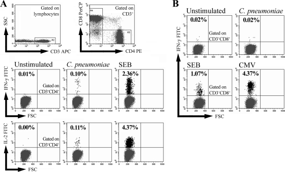
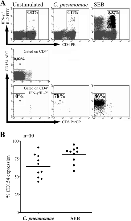
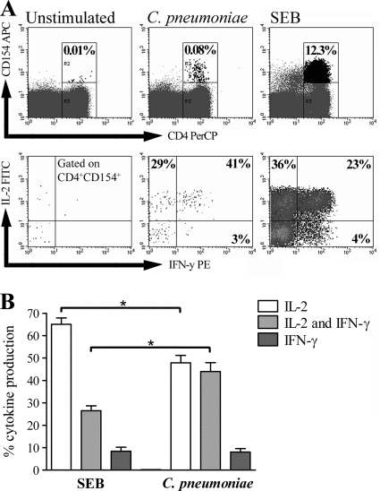
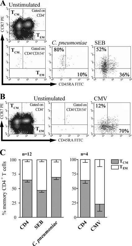
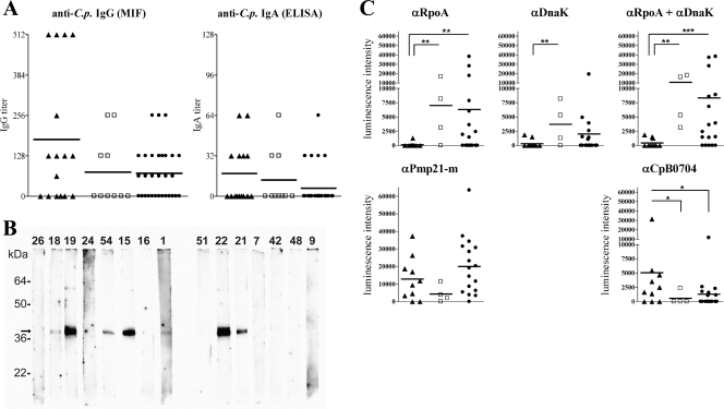
Chlamydia pneumoniae is a frequent pathogen of the respiratory tract, and persistent infections with this obligate intracellular bacterium have been associated with different severe sequelae. Although T-cell activation during acute C. pneumoniae infections has been described, little is known about the frequency or the role of the C. pneumoniae-specific memory T cells that reside in the human body after the resolution of the infection. In the present study, the C. pneumoniae-induced T-cell responses in peripheral blood mononuclear cells of 56 healthy volunteers were analyzed and compared to the donor's serum antibody reactivity toward whole C. pneumoniae as well as recombinant C. pneumoniae antigens. Following short-term stimulation with C. pneumoniae, both gamma interferon (IFN-gamma)- and interleukin-2 (IL-2)-producing CD4(+) T-cell responses could be detected in 16 of 56 healthy individuals. C. pneumoniae-activated CD4(+) T cells expressed CD154, a marker for T-cell receptor-dependent activation, and displayed a phenotype of central memory T cells showing dominant IL-2 production but also IFN-gamma production. Interestingly, individuals with both IFN-gamma- and IL-2-producing responses showed significantly decreased immunoglobulin G reactivity toward C. pneumoniae RpoA and DnaK, antigens known to be strongly upregulated during chlamydial persistence, compared to IgG reactivity of seropositive individuals with no T-cell response or CD4(+) T-cell responses involving the production of a single cytokine (IFN-gamma or IL-2). Our results demonstrate that memory CD4(+) T cells responding to C. pneumoniae stimulation can be detected in the circulation of healthy donors. Furthermore, among seropositive individuals, the presence or the absence of dual IFN-gamma- and IL-2-producing T-cell responses was associated with distinct patterns of antibody responses toward persistence-associated C. pneumoniae antigens.


Figures

FIG. 1.
FIG. 1.
C. pneumoniae-induced T-cell activation in healthy blood donors. The PBMCs of 56 healthy volunteers were stimulated for 6 h with whole C. pneumoniae or control stimuli and assessed for intracellular IFN-γ and IL-2 by flow cytometry. For each donor, 80,000 to 100,000 CD3+ lymphocytes were gated on CD4+ or CD8+ T cells and analyzed for cytokine-positive cells via quadrant analysis. (A) The frequencies of IFN-γ- and IL-2-producing CD3+ CD4+ T cells were determined in the PBMCs of an exemplary donor stimulated with C. pneumoniae or 100 ng SEB or with no stimulation. SSC, side scatter. (B) The frequencies of IFN-γ-producing CD3+ CD8+ T cells in the PBMCs of an exemplary CMV-seropositive donor were determined after stimulation with C. pneumoniae, 100 ng SEB, or CMV-specific peptides.
FIG. 2.
FIG. 2.
CD154 expression among CD4+ T cells activated by C. pneumoniae. The expression of CD154 among activated CD4+ T cells in the PBMCs of donors with C. pneumoniae-induced CD4+ T-cell responses producing IFN-γ and IL-2 was determined after stimulation with C. pneumoniae. As controls, unstimulated PBMCs or PBMCs stimulated with 1 μg SEB were analyzed. For the analysis, 100,000 CD3+ CD4+ T cells were assessed for intracellular IFN-γ and/or IL-2 and were further assessed for the presence of intracellular CD154. (A) The coexpression of CD154 among IFN-γ+ and/or IL-2+ CD4+ T cells following stimulation with C. pneumoniae or SEB is shown for one exemplary donor. CD4+ CD8+ T cells were not analyzed. (B) Proportions of IFN-γ+ and/or IL-2+ CD4+ T cells from 10 different donors coexpressing CD154 after stimulation with C. pneumoniae or SEB. The horizontal lines indicate the mean proportion of CD154 coexpression.
FIG. 3.
FIG. 3.
Cytokine profile of C. pneumoniae-activated CD154+ CD4+ T cells. The distributions of only IL-2+, only IFN-γ+, and both IL-2+ and IFN-γ+ CD154+ CD4+ T cells in the PBMCs of donors with C. pneumoniae-induced CD4+ T cell responses producing IFN-γ and IL-2 were determined after stimulation with C. pneumoniae or SEB. For the analysis, 200,000 CD4+ T cells were measured. (A) The frequencies of only IL-2+, only IFN-γ+, and both IL-2+ and IFN-γ+ CD154+ CD4+ T cells after stimulation with C. pneumoniae or SEB are shown for one exemplary donor. The frequencies of the respective subpopulations were determined by quadrant analysis. (B) Mean frequencies (± SEMs) of only IL-2+, only IFN-γ+, and both IL-2+ and IFN-γ+ CD154+ CD4+ T cells of seven different donors. For comparison, the frequencies of the three subpopulations were normalized to 100% in sum for each donor. Asterisks, significant differences (P < 0.01, tested by ANOVA).
FIG. 4.
FIG. 4.
Memory phenotype analysis of C. pneumoniae-activated CD154+ CD4+ T cells. The levels of expression of CCR7 and CD45RA among total CD4+ T cells and CD154+ CD4+ T cells producing IFN-γ and IL-2 in the PBMCs of donors with C. pneumoniae-induced CD4+ T-cell responses were analyzed after stimulation with C. pneumoniae, SEB, or CMV-specific peptides. For the analysis, 100,000 to 150,000 CD4+ T cells were measured. The quadrants for analysis of CCR7+ and CD45RA TCM and CCR7 and CD45RA+/− TEM were determined for each donor according to the expression of these markers among total CD4+ T cells. (A and B) Proportions of TCM and TEM among CD154+ CD4+ T cells determined after stimulation of PBMCs with C. pneumoniae or SEB (A) or CMV-specific peptides (B) of one exemplary donor and another exemplary CMV-seropositive donor, respectively. (C) Mean frequencies (± SEMs) of TCM and TEM among total CD4+ T cells and CD154+ CD4+ T cells for 12 donors and 4 CMV-seropositive donors stimulated with C. pneumoniae, SEB, or CMV-specific peptides. For comparison, the sum of the frequencies of TCM and TEM was normalized to 100% for each donor.
FIG. 5.
FIG. 5.
Analysis of serum antibody reactivity toward whole C. pneumoniae and different antigens which either are or are not associated with persistent infection. The sera of donors with IFN-γ+ and IL-2+ C. pneumoniae-induced CD4+ T-cell responses (▴), either an IFN-γ+ or an IL-2+ response (□), or no response (•) were analyzed for anti-C. pneumoniae (anti-C.p.) antibodies. *, **, and ***, significant differences representing P values of 0.05, 0.01, and 0.001, respectively (tested by the Kruskal-Wallis test). (A) IgG and IgA antibody titers specific for whole C. pneumoniae elementary bodies were determined in the sera of 56 donors by MIF and ELISA, respectively. The horizontal lines represent the mean titers of each donor group. (B) Strip immunoblots with sera from 15 different exemplary donors obtained by use of the control C. pneumoniae antigen CpB0704. Each number above the lanes represents an individual serum sample, and the arrow indicates the location of CpB0704. (C) Quantitative comparison of chemiluminescence signals of the strip immunoblots for all 31 seropositive donors. The IgG reactivities toward C. pneumoniae antigens RpoA, DnaK, and Pmp21m as well as CpB0704 were determined.

Similar articles

Cited by

References

    1. Apfalter, P. 2006. Chlamydia pneumoniae, stroke, and serological associations: anything learned from the atherosclerosis-cardiovascular literature or do we have to start over again? Stroke 37:756-758. - PubMed
    1. Belland, R. J., S. P. Ouellette, J. Gieffers, and G. I. Byrne. 2004. Chlamydia pneumoniae and atherosclerosis. Cell. Microbiol. 6:117-127. - PubMed
    1. Belland, R. J., G. Zhong, D. D. Crane, D. Hogan, D. Sturdevant, J. Sharma, W. L. Beatty, and H. D. Caldwell. 2003. Genomic transcriptional profiling of the developmental cycle of Chlamydia trachomatis. Proc. Natl. Acad. Sci. U. S. A. 100:8478-8483. - PMC - PubMed
    1. Benagiano, M., A. Azzurri, A. Ciervo, A. Amedei, C. Tamburini, M. Ferrari, J. L. Telford, C. T. Baldari, S. Romagnani, A. Cassone, M. M. D'Elios, and G. Del Prete. 2003. T helper type 1 lymphocytes drive inflammation in human atherosclerotic lesions. Proc. Natl. Acad. Sci. U. S. A. 100:6658-6663. - PMC - PubMed
    1. Benagiano, M., M. M. D'Elios, A. Amedei, A. Azzurri, R. van der Zee, A. Ciervo, G. Rombola, S. Romagnani, A. Cassone, and G. Del Prete. 2005. Human 60-kDa heat shock protein is a target autoantigen of T cells derived from atherosclerotic plaques. J. Immunol. 174:6509-6517. - PubMed

Publication types