Comparative Study
. 2010 May;84(10):5303-13.
doi: 10.1128/JVI.00312-10. Epub 2010 Mar 10.

Sequence variability in clinical and laboratory isolates of herpes simplex virus 1 reveals new mutations

Affiliations
Comparative Study

Sequence variability in clinical and laboratory isolates of herpes simplex virus 1 reveals new mutations

Moriah L Szpara et al. J Virol. 2010 May.

Abstract

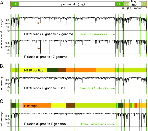
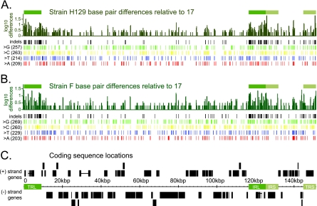
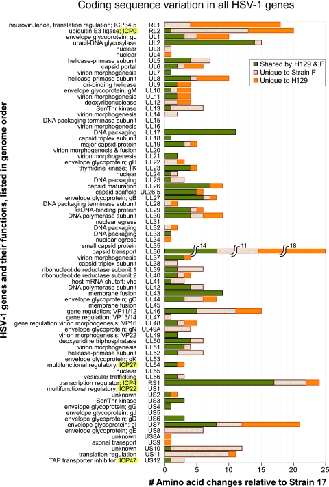
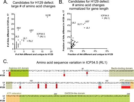
Herpes simplex virus 1 (HSV-1) is a well-adapted human pathogen that can invade the peripheral nervous system and persist there as a lifelong latent infection. Despite their ubiquity, only one natural isolate of HSV-1 (strain 17) has been sequenced. Using Illumina high-throughput sequencing of viral DNA, we obtained the genome sequences of both a laboratory strain (F) and a low-passage clinical isolate (H129). These data demonstrated the extent of interstrain variation across the entire genome of HSV-1 in both coding and noncoding regions. We found many amino acid differences distributed across the proteome of the new strain F sequence and the previously known strain 17, demonstrating the spectrum of variability among wild-type HSV-1 proteins. The clinical isolate, strain H129, displays a unique anterograde spread phenotype for which the causal mutations were completely unknown. We have defined the sequence differences in H129 and propose a number of potentially causal genes, including the neurovirulence protein ICP34.5 (RL1). Further studies will be required to demonstrate which change(s) is sufficient to recapitulate the spread defect of strain H129. Unexpectedly, these data also revealed a frameshift mutation in the UL13 kinase in our strain F isolate, demonstrating how deep genome sequencing can reveal the full complement of background mutations in any given strain, particularly those passaged or plaque purified in a laboratory setting. These data increase our knowledge of sequence variation in large DNA viruses and demonstrate the potential of deep sequencing to yield insight into DNA genome evolution and the variation among different pathogen isolates.


Figures

FIG. 1.
FIG. 1.
Overview of genome coverage for HSV-1 strains F and H129, relative to strain 17 or to their own newly assembled genomes. (A) Diagram of the HSV-1 genome structure, with two unique regions (long and short), each flanked by a pair of terminal repeats. The line graph depicts the depth of sequence read coverage for the newly sequenced strains when each one is aligned to the reference, strain 17. The VNTRs, or reiterations, of strain 17 are overlaid as green lines onto the coverage graphs. Reductions in coverage correlate with the reiterated sequences and also occur more frequently in the terminal repeats that flank each unique region. Brown arrowheads highlight a dip in both new genomes when aligned against strain 17; this is the location of an insertional frameshift in the UL17 gene of strain 17 that leads to alignment mismatch in the nonframeshifted strains F and H129. (B and C) Colored boxes depict the blocks of continuous sequence, or contigs, assembled for each strain. Boxes of the same color represent the same contig; these appear twice when they include sequence in the repeat regions. Sites of reiterations (VNTRs) are overlaid in green. In some cases contigs assembled through a reiteration; in others the contigs terminated at the reiterations. The line graph below the contigs for each strain depicts the improvement in depth of sequence read coverage when data from strain H129 were aligned to the newly assembled H129 genome (B). A similar graph is depicted for strain F (C). Sharp drops in alignment coverage remain at the VNTR sequences inserted from strain 17.
FIG. 2.
FIG. 2.
DNA-level variation across the HSV-1 genome. (A) Bar graph summarizing the number of base pair differences for strain H129 relative to reference strain 17, with differences averaged over a window of 100 bp. The x axis corresponds to the location along the HSV-1 genome (marked in kbp in panel C). The y axis is plotted on a log scale to make small amounts of difference visible on the graph. Green boxes above the bar graph show the location of the terminal repeats, to highlight the increased DNA sequence variation in these areas. Rows of vertical lines depict base pair variations across the genome, with variation separated out by indels (insertions or deletions), and single nucleotide changes. Single base changes are further subdivided by the identity of the base in the new strain: G, C, T, or A. (B) Similar bar graph and statistics for strain F. (C) Location of coding sequences along the genome of HSV-1, which encodes 77 proteins. Plus- and minus-strand-encoded genes are drawn above and below the genome position line. Repeats are denoted by green boxes. Horizontal lines connect the coding sequence boxes of the only spliced transcripts in HSV-1: that of ICP0 (RL2), which is found in the long repeats TRL and IRL, and UL15, which is found around 30 kbp.
FIG. 3.
FIG. 3.
Coding sequence variation across the HSV-1 proteome. Histogram depicting the total number of amino acid differences observed in strains F and H129, relative to the reference strain 17, for every protein in the HSV-1 genome. Changes are color coded to depict the proportion of changes that are different from strain 17 but common between strains F and H129, versus those that are unique to strain F or H129. Proteins are listed in spatial order of occurrence along the HSV-1 genome. Names of the five immediate-early genes, expressed first upon herpesvirus infection of a host cell, are highlighted in yellow (ICP0, ICP27, ICP4, ICP22, and ICP47). Genes in the repeats, ICP34.5 (RL1), ICP0 (RL2), and ICP4 (RS1), are each listed only once, at their first position of occurrence. Ten genes have no amino acid changes at all. Protein functions are summarized to the left of each gene name.
FIG. 4.
FIG. 4.
Potential causative mutations of the H129 spread defect. (A) Scatter plot of the strain H129 proteins, showing the largest overall number of amino acid differences from the wild-type reference strain 17 (y axis) versus the subset of these changes that are also different from the wild-type strain F (x axis) and are thus unique to H129. The most extreme points are labeled to identify the proteins. (B) Because gene length affects the total possible number of observed mutations, the same data were normalized for gene length. The scatter plot presents H129 proteins with the largest overall percentage of amino acids differing from the reference strain 17 (y axis), versus the subset of these changes that are also different from wild-type strain F (x axis). (C) Amino acid alignment of the ICP34.5 (RL1) protein in strains 17, F, and H129. Previously described functional domains are boxed, while amino acid residues that differ from reference strain 17 are highlighted in red. Amino acids that match the reference are represented by dots for clarity; dashes represent deletions in the respective strains.

References

    1. Abaitua, F., R. N. Souto, H. Browne, T. Daikoku, and P. O'Hare. 2009. Characterization of the herpes simplex virus (HSV)-1 tegument protein VP1-2 during infection with the HSV temperature-sensitive mutant tsB7. J. Gen. Virol. 90:2353-2363. - PubMed
    1. Altschul, S. F., W. Gish, W. Miller, E. W. Myers, and D. J. Lipman. 1990. Basic local alignment search tool. J. Mol. Biol. 215:403-410. - PubMed
    1. Archin, N. M., and S. S. Atherton. 2002. Rapid spread of a neurovirulent strain of HSV-1 through the CNS of BALB/c mice following anterior chamber inoculation. J. Neurovirol. 8:122-135. - PubMed
    1. Baird, N. L., P. C. Yeh, R. J. Courtney, and J. W. Wills. 2008. Sequences in the UL11 tegument protein of herpes simplex virus that control association with detergent-resistant membranes. Virology 374:315-321. - PMC - PubMed
    1. Blondeau, C., N. Chbab, C. Beaumont, K. Courvoisier, N. Osterrieder, J. F. Vautherot, and C. Denesvre. 2007. A full UL13 open reading frame in Marek's disease virus (MDV) is dispensable for tumor formation and feather follicle tropism and cannot restore horizontal virus transmission of rRB-1B in vivo. Vet. Res. 38:419-433. - PubMed

Publication types

Associated data