

Glycoprotein D of bovine herpesvirus 5 (BoHV-5) confers an extended host range to BoHV-1 but does not contribute to invasion of the brain
- PMID: 20219909
- PMCID: PMC2876591
- DOI: 10.1128/JVI.00228-10
Glycoprotein D of bovine herpesvirus 5 (BoHV-5) confers an extended host range to BoHV-1 but does not contribute to invasion of the brain
Abstract
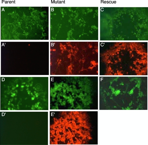
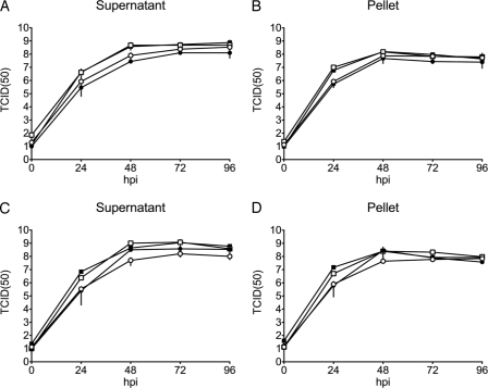
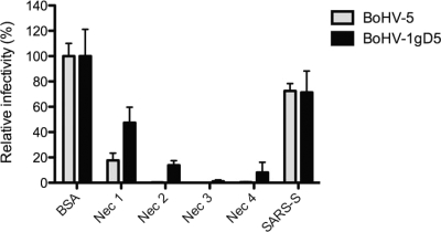
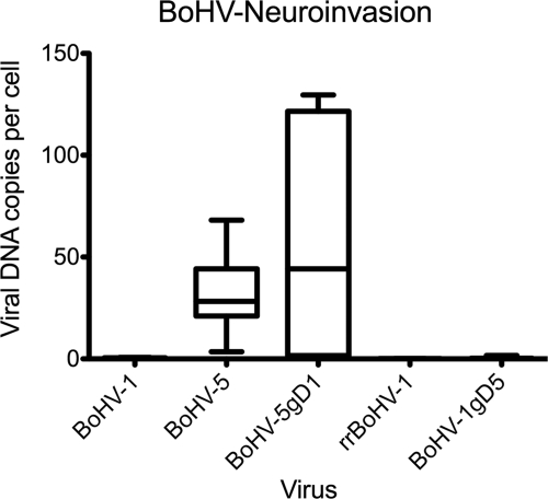
Bovine herpesvirus 1 (BoHV-1) and BoHV-5 are closely related pathogens of cattle, but only BoHV-5 is considered a neuropathogen. We engineered intertypic gD exchange mutants with BoHV-1 and BoHV-5 backbones in order to address their in vitro and in vivo host ranges, with particular interest in invasion of the brain. The new viruses replicated in cell culture with similar dynamics and to titers comparable to those of their wild-type parents. However, gD of BoHV-5 (gD5) was able to interact with a surprisingly broad range of nectins. In vivo, gD5 provided a virulent phenotype to BoHV-1 in AR129 mice, featuring a high incidence of neurological symptoms and early onset of disease. However, only virus with the BoHV-5 backbone, independent of the gD type, was detected in the brain by immunohistology. Thus, gD of BoHV-5 confers an extended cellular host range to BoHV-1 and may be considered a virulence factor but does not contribute to the invasion of the brain.
Figures

Similar articles
-
Molecular and antigenic characterization of Brazilian bovine herpesvirus type 1 isolates recovered from the brain of cattle with neurological disease.Virus Res. 2007 Nov;129(2):191-9. doi: 10.1016/j.virusres.2007.07.014. Epub 2007 Sep 5. Virus Res. 2007. PMID: 17822796
-
Distinctive features of bovine alphaherpesvirus types 1 and 5 and the virus-host interactions that might influence clinical outcomes.Arch Virol. 2020 Feb;165(2):285-301. doi: 10.1007/s00705-019-04494-5. Epub 2019 Dec 16. Arch Virol. 2020. PMID: 31845150 Review.
-
A bovine herpesvirus 5 recombinant defective in the thymidine kinase (TK) gene and a double mutant lacking TK and the glycoprotein E gene are fully attenuated for rabbits.Braz J Med Biol Res. 2010 Feb;43(2):150-9. doi: 10.1590/s0100-879x2009007500030. Epub 2009 Dec 18. Braz J Med Biol Res. 2010. PMID: 20027480
-
Bovine herpesvirus type 5 replication and induction of apoptosis in vitro and in the trigeminal ganglion of experimentally-infected cattle.Comp Immunol Microbiol Infect Dis. 2018 Apr;57:8-14. doi: 10.1016/j.cimid.2018.01.002. Epub 2018 Jan 31. Comp Immunol Microbiol Infect Dis. 2018. PMID: 30017083
-
Biology of bovine herpesvirus 5.Vet J. 2010 May;184(2):138-45. doi: 10.1016/j.tvjl.2009.03.035. Epub 2009 May 5. Vet J. 2010. PMID: 19409823 Review.
Cited by
-
Genetic diversity of 3' region of glycoprotein D gene of bovine herpesvirus 1 and 5.Virus Genes. 2014 Jun;48(3):438-47. doi: 10.1007/s11262-014-1040-5. Epub 2014 Jan 31. Virus Genes. 2014. PMID: 24482291
-
The genome of Chelonid herpesvirus 5 harbors atypical genes.PLoS One. 2012;7(10):e46623. doi: 10.1371/journal.pone.0046623. Epub 2012 Oct 2. PLoS One. 2012. PMID: 23056373 Free PMC article.
-
Bovine herpesvirus glycoprotein D: a review of its structural characteristics and applications in vaccinology.Vet Res. 2014 Oct 31;45(1):111. doi: 10.1186/s13567-014-0111-x. Vet Res. 2014. PMID: 25359626 Free PMC article. Review.
-
Development of an Indirect ELISA for Serological Diagnosis of Bovine herpesvirus 5.PLoS One. 2016 Feb 11;11(2):e0149134. doi: 10.1371/journal.pone.0149134. eCollection 2016. PLoS One. 2016. PMID: 26866923 Free PMC article.
References
-
- Abdelmagid, O. Y., H. C. Minocha, J. K. Collins, and S. I. Chowdhury. 1995. Fine mapping of bovine herpesvirus-1 (BHV-1) glycoprotein D (gD) neutralizing epitopes by type-specific monoclonal antibodies and sequence comparison with BHV-5 gD. Virology 206:242-253. - PubMed
-
- Al-Mubarak, A., and S. I. Chowdhury. 2004. In the absence of glycoprotein I (gI), gE determines bovine herpesvirus type 5 neuroinvasiveness and neurovirulence. J. Neurovirol. 10:233-243. - PubMed
-
- Ashbaugh, S. E., K. E. Thompson, E. B. Belknap, P. C. Schultheiss, S. Chowdhury, and J. K. Collins. 1997. Specific detection of shedding and latency of bovine herpesvirus 1 and 5 using a nested polymerase chain reaction. J. Vet. Diagn. Invest. 9:387-394. - PubMed
Publication types
MeSH terms
Substances
LinkOut - more resources
Full Text Sources
