. 2010 May;84(10):5181-90.
doi: 10.1128/JVI.02382-09. Epub 2010 Mar 10.

Role of human immunodeficiency virus type 1 integrase in uncoating of the viral core

Affiliations

Role of human immunodeficiency virus type 1 integrase in uncoating of the viral core

Marisa S Briones et al. J Virol. 2010 May.

Abstract

After membrane fusion with a target cell, the core of human immunodeficiency virus type 1 (HIV-1) enters into the cytoplasm, where uncoating occurs. The cone-shaped core is composed of the viral capsid protein (CA), which disassembles during uncoating. The underlying factors and mechanisms governing uncoating are poorly understood. Several CA mutations can cause changes in core stability and a block at reverse transcription, demonstrating the requirement for optimal core stability during viral replication. HIV-1 integrase (IN) catalyzes the insertion of the viral cDNA into the host genome, and certain IN mutations are pleiotropic. Similar to some CA mutants, two IN mutants, one with a complete deletion of IN (NL-DeltaIN) and the other with a Cys-to-Ser substitution (NL-C130S), were noninfectious, with a replication block at reverse transcription. Compared to the wild type (WT), the cytoplasmic CA levels of the IN mutants in infected cells were reduced, suggesting accelerated uncoating. The role of IN during uncoating was examined by isolating and characterizing cores from NL-DeltaIN and NL-C130S. Both IN mutants could form functional cores, but the core yield and stability were decreased. Also, virion incorporation of cyclophilin A (CypA), a cellular peptidyl-prolyl isomerase that binds specifically to CA, was decreased in the IN mutants. Cores isolated from WT virus depleted of CypA had an unstable-core phenotype, confirming a role of CypA in promoting optimal core stability. Taken together, our results indicate that IN is required during uncoating for maintaining CypA-CA interaction, which promotes optimal stability of the viral core.


Figures

FIG. 1.
FIG. 1.
trans incorporation of IN rescues the reverse transcription defect of the IN mutants. 293T cells were transfected with pNL-C130S or pNL-ΔIN or cotransfected with pNL-C130S and pLR2P-Vpr-IN, or pNL-ΔIN and pLR2P-Vpr-IN. WT NL4-3 was used as a positive control. Two days posttransfection, viruses were collected from the culture media and equal amounts of p24-equivalent of each virus were used to infect CEM-GFP cells. Quantitative analysis of viral cDNA synthesis was done by real-time PCR. The amount of early reverse-transcribed viral DNA present in infected cells was measured 16 h after infection with WT, NL-C130S, NL-C130S plus Vpr-IN, NL-ΔIN, or NL-ΔIN plus Vpr-IN viruses. Control infections with culture medium alone (Mock) and cells infected with heat-inactivated WT virus (HI) were used as negative controls. The values are −sssDNA copy numbers and are expressed as the average percentages of WT virus from two experiments.
FIG. 2.
FIG. 2.
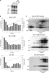
Isolation of viral cores from WT HIV-1 and IN mutant viruses. (A) Western blot of concentrated viruses. WT and IN mutant viruses prepared by transient transfections of 293T cells were concentrated over a 20% sucrose cushion, and the viral titer was measured by p24 ELISA. Fifty nanograms of p24-equivalent concentrated virus was separated on a 10% SDS-polyacrylamide gel, and viral protein expression was examined by Western blot analysis. The blots were probed with human anti-HIV serum. (B) Graphic depiction of the p24 contents obtained by p24 ELISA (bars) and the densities of the sucrose gradient fractions (lines) for all three viruses. (C) Western blots of gradient fractions after ultracentrifugation. Each blot was probed with human anti-HIV serum. Fractions containing cores are indicated. In panels A and C, viral proteins are labeled as reverse transcriptase (RT), integrase (IN), capsid (CA), and matrix (MA).
FIG. 3.
FIG. 3.
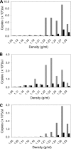
Viral cDNA synthesis of gradient fractions from WT and IN mutant viruses. Fraction samples from WT (A) and both IN mutant viruses, NL-C130S (B) and NL-ΔIN (C), were subjected to an endogenous reverse-transcription assay. After 16 h, each sample was analyzed for the early reverse transcription product using real-time PCR with primers specific for −sssDNA (gray bars). As a control, efavirenz was added to the reaction mixture to a final concentration of 7.5 μM (black bars). The results are expressed as copy numbers per microliter for each fraction with the indicated densities and are representative of at least three independent experiments for each virus.
FIG. 4.
FIG. 4.
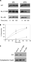
CA disassembly of WT and IN mutant viruses in vitro and in vivo. (A) Western blots illustrating disassembly of CA from the viral core. Gradient fractions corresponding to viral cores were pooled and placed at either 4°C or 37°C for 0, 60, or 120 min. Samples were then subjected to ultracentrifugation at 100,000 × g for 20 min at 4°C. The pellets were resuspended in 2% SDS sample buffer for Western blot analysis, and the blots were probed with a monoclonal anti-CA antibody. The blots were analyzed by chemiluminescence of the horseradish peroxidase (HRP)-conjugated secondary antibody and are representative of at least 3 independent experiments. (B) Disassembly kinetics of viral cores at 37°C. The Western blots shown in panel A were scanned, and ImageQuant TL software (GE Healthcare) was used to quantify band intensity. Percent disassembly was determined by using the value at time zero as the total p24 amount. The results are the mean values ± standard errors of the mean (SEM) of at least three independent experiments for WT (▴), NL-C130S (▪), and NL-ΔIN (⧫). (C) CA degradation in the cytoplasmic cell lysates of infected cells. HeLa CD4+ cells were infected with either WT, NL-C130S, or NL-ΔIN virus for 6 h at 37°C. The CA contents in cytoplasmic lysates were examined by Western blot analysis, and the blots were probed with polyclonal anti-CA antibodies (top). The cytoplasmic CypA content, probed with polyclonal anti-CypA antibody, served as a specificity control and was used to normalize the amount of lysate loaded onto the gel (bottom). The results are representative of two independent experiments.
FIG. 5.
FIG. 5.
Decrease in CypA incorporation into IN mutant virus. WT and IN mutant viruses prepared by transient transfections of 293T cells were concentrated over a 20% sucrose cushion, and the viral titer was measured by p24 ELISA. Fifty nanograms of p24-equivalent concentrated virus was separated on a 10% SDS-polyacrylamide gel, and Western blot analysis was carried out using a polyclonal anti-CypA antibody to measure viral incorporation of CypA (top) and a monoclonal antibody to CA (bottom).
FIG. 6.
FIG. 6.
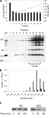
Depletion of CypA from HIV-1 results in an unstable-core phenotype. (A) Isolation of viral cores from WT virus depleted of CypA. (Top) Graphic depiction of the p24 contents obtained by p24 ELISA (bars) and the densities of the sucrose gradient fractions (gray lines). (Bottom) Western blot of gradient fractions after ultracentrifugation that was probed with human anti-HIV serum. Fifty nanograms of p24-equivalent concentrated virus (CV) was added as a size marker for viral proteins. The other symbols have the same meaning as in Fig. 2. (B) Viral cDNA synthesis from gradient fractions. The ERT assay was used to measure −sssDNA by real-time PCR in the absence (gray bars) or presence (black bars) of 7.5 μM efavirenz. The results are expressed as the copy number per microliter for each fraction with the indicated densities. (C) In vitro core disassembly assay. Isolated cores were pooled and placed at either 4°C or 37°C for up to 120 min. Samples were subjected to ultracentrifugation, and the CA contents of the pellet fractions were determined by Western blot analysis by probing with monoclonal anti-CA antibody. In all panels, the results are representative of at least three independent experiments.

Similar articles

Cited by

References

    1. Accola, M. A., A. Ohagen, and H. G. Gottlinger. 2000. Isolation of human immunodeficiency virus type 1 cores: retention of Vpr in the absence of p6(gag). J. Virol. 74:6198-6202. - PMC - PubMed
    1. Adamson, C. S., K. Salzwedel, and E. O. Freed. 2009. Virus maturation as a new HIV-1 therapeutic target. Exp. Opin. Ther. Targets 13:895-908. - PMC - PubMed
    1. Aiken, C. 1997. Pseudotyping human immunodeficiency virus type 1 (HIV-1) by the glycoprotein of vesicular stomatitis virus targets HIV-1 entry to an endocytic pathway and suppresses both the requirement for Nef and the sensitivity to cyclosporin A. J. Virol. 71:5871-5877. - PMC - PubMed
    1. Aiken, C. 2006. Viral and cellular factors that regulate HIV-1 uncoating. Curr. Opin. HIV AIDS 1:194-199. - PubMed
    1. Auewarakul, P., P. Wacharapornin, S. Srichatrapimuk, S. Chutipongtanate, and P. Puthavathana. 2005. Uncoating of HIV-1 requires cellular activation. Virology 337:93-101. - PubMed

Publication types

Substances

LinkOut - more resources