

doi: 10.1128/JVI.00098-10.
Epub 2010 Mar 10.
Visualizing viral dissemination in the mouse nervous system, using a green fluorescent protein-expressing Borna disease virus vector
Affiliations
- PMID: 20219925
- PMCID: PMC2863799
- DOI: 10.1128/JVI.00098-10
Item in Clipboard
Visualizing viral dissemination in the mouse nervous system, using a green fluorescent protein-expressing Borna disease virus vector
J Virol.
2010 May.
Abstract
Borna disease virus (BDV) frequently persists in the brain of infected animals. To analyze viral dissemination in the mouse nervous system, we generated a mouse-adapted virus that expresses green fluorescent protein (GFP). This viral vector supported GFP expression for up to 150 days and possessed an extraordinary staining capacity, visualizing complete dendritic arbors as well as individual axonal fibers of infected neurons. GFP-positive cells were first detected in cortical areas from where the virus disseminated through the entire central nervous system (CNS). Late in infection, GFP expression was found in the sciatic nerve, demonstrating viral spread from the central to the peripheral nervous system.
Figures
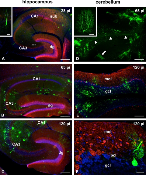
Distribution of mBDV-GFP-infected cells in the hippocampus and the cerebellum. C57BL/6 mice were infected with 10,000 FFU of mBDV-GFP. At the indicated time points (days) p.i., two animals were sacrificed for analysis. (A) Survey of an anticalbindin-immunostained hippocampal section showing the typical calbindin-immunoreactive labeling of granule cells, their mossy fiber projection (mf), and CA1 pyramidal neurons at 28 days p.i. Hoechst nuclear staining illustrates the cytoarchitectural organization of the hippocampus. In the subiculum (sub) GFP-containing cells, presumably astrocytes, are observed. In the CA3 region a single mBDV-GFP-infected pyramidal neuron can be identified. Strong GFP expression allows the visualization of the dendritic tree of this neuron (inset). dg, dentate gyrus. (B) At 65 days p.i. the infection had spread throughout the hippocampus. mBDV-GFP-infected neurons were found in the CA3 region and the granule cell layer (green dots) of the dentate gyrus. (C) At 120 days p.i. the intensity of GFP expression had increased and dendritic processes of infected neurons were easily detected in the CA3 region. The cytoarchitecture of the hippocampus and the calbindin staining of the dentate gyrus appeared unaltered. (D) Overview of cerebellar folia showing strong GFP labeling in the granular cell layer and of axons in the white matter (arrowheads) at 65 days p.i. The arrow points to a cluster of infected Purkinje cells (PC) extending their dendrites into the molecular layer. The inset shows the complete dendritic arbor of an infected PC at high magnification. (E) Micrograph of a cerebellar folium immunolabeled with an antibody against parvalbumin (PARV), a specific marker for PC, at 120 days p.i. Counterstaining with Hoechst stain reveals normal cytoarchitecture. Some BDV-infected PC were found between PARV-positive PC. In the granular layer (blue) many infected cells and their processes were visible. gcl, granular cell layer; mol, molecular layer. (F) An example of an mBDV-GFP-infected PC with its dendrites. The cell body is neighbored by PARV-immunoreactive somata of GFP-negative Purkinje neurons. pcl, Purkinje cell layer. Bars: 250 μm (A), 15 μm (inset of panel A), 250 μm (B and C), 600 μm (D), 60 μm (inset of panel D), 200 μm (E), and 30 μm (F).
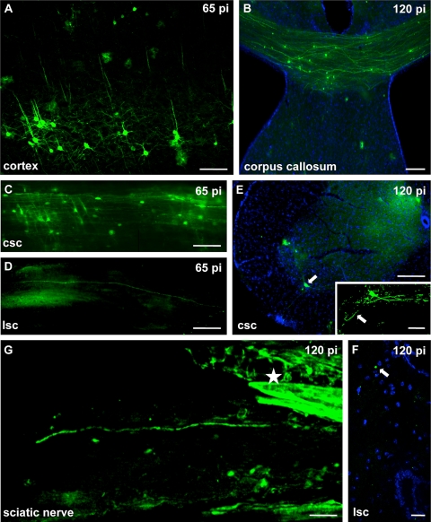
mBDV-GFP infection of the cortex, the spinal cord, and the peripheral nervous system. C57BL/6 mice were infected with 10,000 FFU of mBDV-GFP. At the indicated time points p.i., two animals were sacrificed for analysis. (A) Intensely GFP-labeled cortical neurons with their typical dendritic pattern. (B) Coronal section of the corpus callosum reveals many GFP-positive axon profiles at 120 days p.i. (C) At 65 days p.i. numerous GFP-labeled cell bodies and axonal profiles were visible in this longitudinal section of the cervical (csc) part of the spinal cord. (D) At that time point, a single axon protruding into the lumbar (lsc) portion of the spinal cord could be traced. (E) Cross section through the cervical spinal cord exhibiting an infected motor neuron (arrow) in the ventral horn. For orientation the section was stained with Hoechst stain. The higher-magnification inset shows the GFP-positive motor neuron, illustrating the dendrites and the efferent axon (arrow) directed toward the anterior radix of the spinal cord. (F) Cross section of the lumbar spinal cord. At a very late time point (120 days p.i.) GFP-labeled circular structures (arrow), presumably representing axons, were found in the posterior part of the lumbar spinal cord. (G) GFP-positive nerve fiber of the peripheral sciatic nerve and the strongly GFP-positive perineural sheaths (asterisks). Bars: 100 μm (A), 50 μm (B), 200 μm (C and D), 250 μm (E), 50 μm (inset of panel E), 25 μm (F), 100 μm (G).
Loss of GFP expression at late time points after infection. C57BL/6 mice were infected with 10,000 FFU of mBDV-GFP. At the indicated time points p.i., the animals were sacrificed for analysis. (A) Analyses of virus load and GFP expression by Western blotting using a rabbit antiserum specific for BDV-N and a monoclonal antibody against GFP (Stratagene). Correct loading of the gel was verified by staining with a monoclonal antibody against β-tubulin (Sigma). (B) Northern blot analyses of 5 μg (NPX) and 20 μg (GFP) of total RNA isolated from the brains of two animals (lanes A and B) sacrificed 10 weeks p.i. The analyses were performed as previously described (12). The identities of viral transcripts are indicated. For a loading control, we show the ethidium bromide staining of the cellular 18S rRNA. (C) Staining of the dentate gyrus (DG) of an infected mouse at 150 days p.i. with a rabbit antiserum specific for BDV-N. Bound antibodies were detected with a Cy3-conjugated goat anti-rabbit IgG (red). The granule cell layer is intact as shown by Hoechst nuclear staining. Bar, 100 μm. (D) Schematic representation of the regulatory sequences that govern the transcription of the GFP gene. The position of the primer used for sequencing is indicated. (E) Sequence analyses of RT-PCR fragments amplified from viral RNA isolated from mice 4 and 10 weeks after infection with mBDV-GFP, respectively. The electropherograms represent the results of bulk sequencing of the PCR fragments. The selected windows show the transcription regulatory sequences indicated in panel D. Below are shown the sequencing results of individual PCR fragments subcloned into pTopo (Invitrogen). Nucleotides that diverge from the wild-type sequence are shown in red.
Similar articles
-
Neurological diseases and viral dynamics in the brains of neonatally borna disease virus-infected gerbils.Virology. 2001 Mar 30;282(1):65-76. doi: 10.1006/viro.2001.0813. Virology. 2001. PMID: 11259191
-
[The neuropathogenesis of Borna disease virus infection].Nihon Rinsho. 2001 Aug;59(8):1605-13. Nihon Rinsho. 2001. PMID: 11519168 Review. Japanese.
-
Bornavirus and the brain.J Infect Dis. 2002 Dec 1;186 Suppl 2:S241-7. doi: 10.1086/344936. J Infect Dis. 2002. PMID: 12424704 Review.
-
Generation of a non-transmissive Borna disease virus vector lacking both matrix and glycoprotein genes.Microbiol Immunol. 2017 Sep;61(9):380-386. doi: 10.1111/1348-0421.12505. Microbiol Immunol. 2017. PMID: 28776750
-
Rabies and borna disease. A comparative pathogenetic study of two neurovirulent agents.Lab Invest. 1993 Mar;68(3):285-95. Lab Invest. 1993. PMID: 8450648
Cited by
-
Avian Bornavirus Research-A Comprehensive Review.Viruses. 2022 Jul 11;14(7):1513. doi: 10.3390/v14071513. Viruses. 2022. PMID: 35891493 Free PMC article. Review.
-
Genetic stability of the open reading frame 2 (ORF2) of borna disease virus 1 (BoDV-1) distributed in cattle in Hokkaido.J Vet Med Sci. 2021 Oct 2;83(10):1526-1533. doi: 10.1292/jvms.21-0155. Epub 2021 Aug 13. J Vet Med Sci. 2021. PMID: 34393150 Free PMC article.
-
Word recognition memory and serum levels of Borna disease virus specific circulating immune complexes in obsessive-compulsive disorder.BMC Psychiatry. 2022 Sep 8;22(1):597. doi: 10.1186/s12888-022-04208-3. BMC Psychiatry. 2022. PMID: 36076225 Free PMC article.
-
A novel borna disease virus vector system that stably expresses foreign proteins from an intercistronic noncoding region.J Virol. 2011 Dec;85(23):12170-8. doi: 10.1128/JVI.05554-11. Epub 2011 Sep 21. J Virol. 2011. PMID: 21937656 Free PMC article.
-
A novel porcine reproductive and respiratory syndrome virus vector system that stably expresses enhanced green fluorescent protein as a separate transcription unit.Vet Res. 2013 Oct 31;44(1):104. doi: 10.1186/1297-9716-44-104. Vet Res. 2013. PMID: 24176053 Free PMC article.
References
-
- Celio, M. R. 1990. Calbindin D-28k and parvalbumin in the rat nervous system. Neuroscience 35:375-475. - PubMed
-
- de la Torre, J. C. 2002. Bornavirus and the brain. J. Infect. Dis. 186(Suppl. 2):S241-S247. - PubMed
-
- Herzog, S., C. Kompter, K. Frese, and R. Rott. 1984. Replication of Borna disease virus in rats: age-dependent differences in tissue distribution. Med. Microbiol. Immunol. 173:171-177. - PubMed
Publication types
MeSH terms
Substances
LinkOut - more resources
Full Text Sources
