. 2010 May;84(10):4889-97.
doi: 10.1128/JVI.00057-10. Epub 2010 Mar 10.

Electron cryotomography of Tula hantavirus suggests a unique assembly paradigm for enveloped viruses

Affiliations

Electron cryotomography of Tula hantavirus suggests a unique assembly paradigm for enveloped viruses

Juha T Huiskonen et al. J Virol. 2010 May.

Abstract

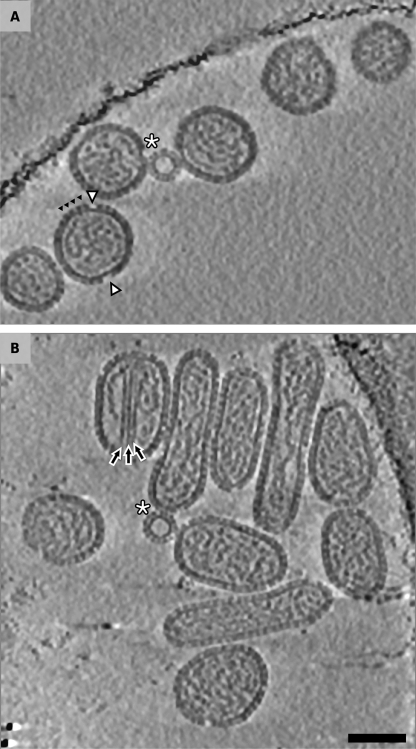
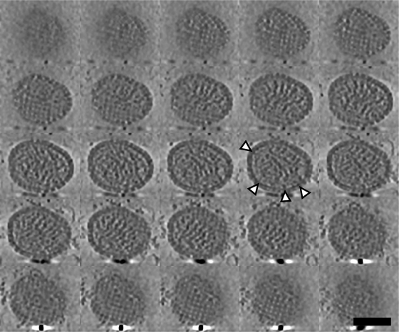
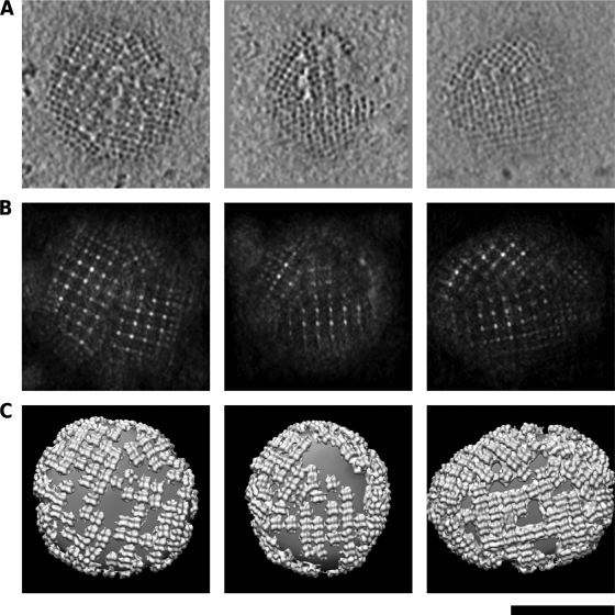
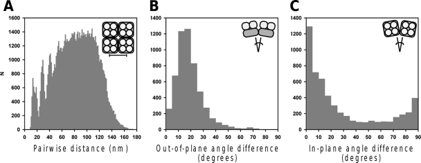
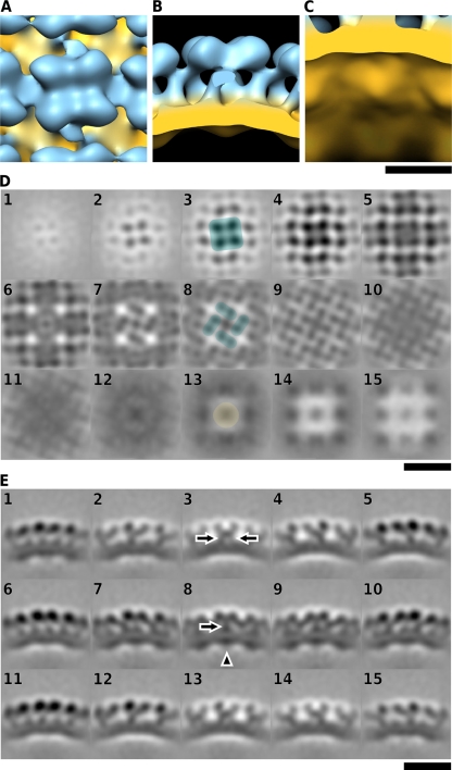
Hantaviruses (family Bunyaviridae) are rodent-borne emerging viruses that cause a serious, worldwide threat to human health. Hantavirus diseases include hemorrhagic fever with renal syndrome and hantavirus cardiopulmonary syndrome. Virions are enveloped and contain a tripartite single-stranded negative-sense RNA genome. Two types of glycoproteins, G(N) and G(C), are embedded in the viral membrane and form protrusions, or "spikes." The membrane encloses a ribonucleoprotein core, which consists of the RNA segments, the nucleocapsid protein, and the RNA-dependent RNA polymerase. Detailed information on hantavirus virion structure and glycoprotein spike composition is scarce. Here, we have studied the structures of Tula hantavirus virions using electron cryomicroscopy and tomography. Three-dimensional density maps show how the hantavirus surface glycoproteins, membrane, and ribonucleoprotein are organized. The structure of the G(N)-G(C) spike complex was solved to 3.6-nm resolution by averaging tomographic subvolumes. Each spike complex is a square-shaped assembly with 4-fold symmetry. Spike complexes formed ordered patches on the viral membrane by means of specific lateral interactions. These interactions may be sufficient for creating membrane curvature during virus budding. In conclusion, the structure and assembly principles of Tula hantavirus exemplify a unique assembly paradigm for enveloped viruses.


Figures

FIG. 1.
FIG. 1.
Morphology of Tula hantavirus particles. Computational slices (5.4 nm thick) of the tomographic reconstructions. The slice cuts approximately through the center of the particles. The shape of the particles varied from tubular to spherical. Internal densities including the membrane and RNPs were resolved in the tomographic reconstruction. Some spikes (filled triangles) and surface areas devoid of spikes (arrowheads) are indicated in panel A. Small spherical particles devoid of any RNP density are indicated with asterisks. Straight, rod-shaped densities distinct from the RNP densities are indicated with arrows in panel B. The reconstructions have been denoised using nonisotropic diffusion. Scale bar, 50 nm.
FIG. 2.
FIG. 2.
Organization of the ribonucleoprotein. Consecutive computational slices (2.7 nm thick) through a tomographic reconstruction of one virion. The reconstruction has been denoised using nonisotropic diffusion. The RNP particles appear as 8-nm-wide threads. Some RNP-membrane connections are indicated. Scale bar, 100 nm.
FIG. 3.
FIG. 3.
Glycoprotein spikes form ordered patches on the viral membrane. (A) Computational slices (5.4 nm thick) through three individual denoised virion reconstructions. Four-lobed spikes are often evident and create ordered patches. Between the patches, exposed areas of the membrane are present. (B) Cross sections (5.4 nm thick) through the maximum cross-correlation maps after correlation-based template matching. Cross-correlation was calculated between the virion reconstructions in panel A and a model of the glycoprotein spike. (C) Surface models of the spike lattices. The model of the glycoprotein spike was placed into the positions with high cross-correlation values shown in panel B. The smooth surface represents the viral membrane. Scale bar, 100 nm (A and C).
FIG. 4.
FIG. 4.
Quantitative analysis of the glycoprotein spike patches. (A) Histogram of all pairwise distances calculated within each virion. (B) Histogram of all differences in out-of-plane angles calculated between all pairs of neighboring spikes. (C) Histogram of all differences in in-plane angles calculated between all pairs of neighboring spikes. The insets illustrate the performed measurement in each case.
FIG. 5.
FIG. 5.
Averaged structure of the glycoprotein spike complex. (A to C) The average spike structure is shown from the top (A), from the side (B), and at an angle from inside the virion (C). Surface was rendered at 0.6σ above the mean density. The molecular mass confined by this surface was 480 kDa for the ectodomain, as determined in Bsoft (14). Scale bar, 10 nm. (D) Slices (1.3 nm thick) through the volume from the top, tangential to the membrane. The four globular domains forming the membrane-distal part of one spike (slice 3), the pairs of peripheral stalks forming spike-to-spike interfaces (slice 8), and the additional density in the cytoplasmic side of the membrane (slice 13) are colored. (E) Slices (1.3 nm thick) through the volume from the side. Peripheral stalks (slice 3) and a central stalk (slice 8) are indicated with arrows; intraviral density is indicated with an arrowhead. Scale bar, 20 nm (D and E).

Similar articles

Cited by

References

    1. Adrian, M., J. Dubochet, J. Lepault, and A. W. McDowall. 1984. Cryo-electron microscopy of viruses. Nature 308:32-36. - PubMed
    1. Baker, T. S., N. H. Olson, and S. D. Fuller. 1999. Adding the third dimension to virus life cycles: three-dimensional reconstruction of icosahedral viruses from cryo-electron micrographs. Microbiol. Mol. Biol. Rev. 63:862-922. - PMC - PubMed
    1. Butan, C., D. C. Winkler, J. B. Heymann, R. C. Craven, and A. C. Steven. 2008. RSV capsid polymorphism correlates with polymerization efficiency and envelope glycoprotein content: implications that nucleation controls morphogenesis. J. Mol. Biol. 376:1168-1181. - PMC - PubMed
    1. Cardone, G., J. G. Purdy, N. Cheng, R. C. Craven, and A. C. Steven. 2009. Visualization of a missing link in retrovirus capsid assembly. Nature 457:694-698. - PMC - PubMed
    1. Deyde, V. M., A. A. Rizvanov, J. Chase, E. W. Otteson, and S. C. St. Jeor. 2005. Interactions and trafficking of Andes and Sin Nombre Hantavirus glycoproteins G1 and G2. Virology 331:307-315. - PubMed

Publication types

LinkOut - more resources