. 2010 Jun;298(6):C1538-48.
doi: 10.1152/ajpcell.00467.2009. Epub 2010 Mar 10.

Apolipoprotein A-I mimetic 4F alters the function of human monocyte-derived macrophages

Affiliations

Apolipoprotein A-I mimetic 4F alters the function of human monocyte-derived macrophages

Lesley E Smythies et al. Am J Physiol Cell Physiol. 2010 Jun.

Abstract

HDL and its major protein component apolipoprotein A-I (apoA-I) exert anti-inflammatory effects, inhibit monocyte chemotaxis/adhesion, and reduce vascular macrophage content in inflammatory conditions. In this study, we tested the hypothesis that the apoA-I mimetic 4F modulates the function of monocyte-derived macrophages (MDMs) by regulating the expression of key cell surface receptors on MDMs. Primary human monocytes and THP-1 cells were treated with 4F, apoA-I, or vehicle for 7 days and analyzed for expression of cell surface markers, adhesion to human endothelial cells, phagocytic function, cholesterol efflux capacity, and lipid raft organization. 4F and apoA-I treatment decreased the expression of HLA-DR, CD86, CD11b, CD11c, CD14, and Toll-like receptor-4 (TLR-4) compared with control cells, suggesting the induction of monocyte differentiation. Both treatments abolished LPS-induced mRNA for monocyte chemotactic protein-1 (MCP-1), macrophage inflammatory protein-1 (MIP-1), regulated on activation, normal T-expressed and presumably secreted (RANTES), IL-6, and TNF-alpha but significantly upregulated LPS-induced IL-10 expression. Moreover, 4F and apoA-I induced a 90% reduction in the expression of CD49d, a ligand for the VCAM-1 receptor, with a concurrent decrease in monocyte adhesion (55% reduction) to human endothelial cells and transendothelial migration (34 and 27% for 4F and apoA-I treatments) compared with vehicle treatment. In addition, phagocytosis of dextran-FITC beads was inhibited by 4F and apoA-I, a response associated with reduced expression of CD32. Finally, 4F and apoA-I stimulated cholesterol efflux from MDMs, leading to cholesterol depletion and disruption of lipid rafts. These data provide evidence that 4F, similar to apoA-I, induces profound functional changes in MDMs, possibly due to differentiation to an anti-inflammatory phenotype.


Figures
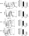
Fig. 1.
Fig. 1.
4F and apoliprotein (apo)A-I alter macrophage phenotype. Primary human monocytes were treated with 4F (50 μg/106 cells), apoA-I (50 μg/106 cells), or vehicle control for 7 days. Cells were stained with antibodies to HLA-DR, CD86, CD11b, and CD11c, and expression of these surface markers was monitored by flow cytometry. Left: monocyte-derived macrophage (MDM) surface marker expression, depicted as a representative histogram for each marker under different treatment conditions. Hatched gray lines, control cells; solid black lines, cells + 4F; dashed lines, cells + apoA-I. Numbers depict the mean fluorescence intensity of the corresponding histogram. Right: changes in percentage of cells expressing each marker. Percentage of cells expressing a particular marker is calculated with respect to control cells, designated 100%. Data are means ± SE; n = 5. *P < 0.05, significant difference between control samples and treatment groups.
Fig. 2.
Fig. 2.
4F enhances IL-10 mRNA expression in MDMs. Primary human monocytes were pretreated with 4F (50 μg/106 cells), apoA-I (50 μg/106 cells), or vehicle for 7 days. 4F induces a 2-fold upregulation of IL-10 with no significant effect on the other genes, whereas apoA-I induces an upregulation of IL-6 (4.5-fold) and IL-10 (2-fold). Data are means ± SE; n = 5.
Fig. 3.
Fig. 3.
4F abolishes LPS-induced inflammatory chemokine and cytokine mRNA upregulation and enhances IL-10 mRNA expression. Primary human monocytes were pretreated with 4F (50 μg/106 cells), apoA-I (50 μg/106 cells), or vehicle for 7 days stimulated with LPS (1.0 μg/ml) for 18 h. Treatment with 4F, apoA-I, or 4F + apoA-I significantly decreased LPS-induced monocyte chemotactic protein-1 (MCP-1), macrophage inflammatory protein-1 (MIP-1), regulated on activation normal T-expressed and presumably secreted (RANTES), IL-6, and TNF-α and enhanced IL-10 mRNA. Data are means ± SE; n = 5. *P < 0.05, significant difference between LPS-treated and 4F-, apoA-I-, or 4F + apoA-I-treated cells.
Fig. 4.
Fig. 4.
4F attenuates Toll-like receptor-4 (TLR-4) and CD14 expression. Pretreatment of MDMs with 4F (50 μg/106 cells) or apoA-I (50 μg/106 cells) for 7 days downregulates surface CD14 and TLR-4 compared with vehicle control as measured by flow cytometry. Left: MDM surface marker expression, depicted as a representative histogram for each marker under different treatment conditions. Hatched gray lines, control cells; solid black lines, cells + 4F; dashed lines, cells + apoA-I. Numbers depict the mean fluorescence intensity of the corresponding histogram. Right: changes in percentage of cells expressing the markers. Percentage of cells expressing a particular marker is calculated with respect to control cells, designated 100%. Data are means ± SE; n = 5. *P < 0.05, significant difference between control samples and treated cells.
Fig. 5.
Fig. 5.
4F inhibits monocyte adhesion to and transmigration through human umbilical vein endothelial cells (HUVECs). A: MDMs were treated with 4F (50 μg/106 cells), apoA-I (50 ug/106 cells), or vehicle control for 7 days and loaded with calcein and their adhesion to LPS-stimulated HUVECs was measured. Data are means ± SE. *P < 0.001, 4F and apoA-I vs. control. B: MDMs pretreated with 4F and apoA-I express decreased levels of CD49d compared with control-treated cells. Numbers depict the mean fluorescence intensity of the corresponding histogram. Percentage of cells expressing a particular marker is calculated with respect to control cells, designated 100%. Hatched gray lines, control cells; solid black lines, cells + 4F; dashed lines, cells + apoA-I. C: pretreatment of MDMs with 4F (50 μg/106 cells), apoA-I (50 ug/106 cells), or vehicle control for 7 days inhibits transendothelial migration. HUVECs were plated on Transwell filters, and calcein-loaded MDMs pretreated with vehicle, 4F, or apoA-I were added to the filters. Fluorescence of the migrated cells (measured in the lower chamber) is expressed as a percentage of cells loaded on the filter. Transendothelial migration of 4F-treated cells is 34% less and apoA-I treated cells is 27% less than controls. Data are means ± SE; n = 6. *P < 0.01, compared with controls.
Fig. 6.
Fig. 6.
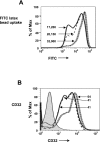
4F and apoA-I decrease the phagocytic activity of macrophages. Pretreatment of MDMs with 4F (50 μg/106 cells), apoA-I (50 ug/106 cells), or vehicle control for 7 days inhibits phagocytosis of FITC-labeled latex beads as indicated by reduced mean fluorescence intensity (A) and decreases the expression of CD32 (B). Numbers depict the mean fluorescence intensity of the corresponding histogram. Hatched gray lines, control cells; solid black lines, cells + 4F; dashed lines, cells + apoA-I.
Fig. 7.
Fig. 7.
4F stimulates cholesterol efflux and decreases membrane-associated rafts. A: primary human monocytes were loaded with [14C]cholesterol and treated with 4F + apoA-I for 18 h. Cholesterol effluxed from the cell was measured and expressed as a percentage of total cholesterol (cell + medium). Black bars, vehicle; open bars, cells + 4F; hatched bars, cells + apoA-I . Data are means ± SE; n = 6. *P < 0.001, significant difference between control samples and treated cells. B: cells were treated with medium alone, 4F (50 μg/106 cells), or apoA-I (50 ug/106 cells) for 4 days and stained for lipid rafts and nuclei using Alexa fluor 488-CT-B conjugate (FITC) and DAPI, respectively. C: number of cells expressing lipid rafts, determined by ImageJ analysis, and expressed as rafts/nuclei, was significantly reduced in cells treated with 4F and apoA-I compared with medium alone (5.6 ± 2.3, 6.4 ± 4.6, and 18.4 ± 5.6, respectively). *P < 0.008. Data represent 4 experiments.

Similar articles

Cited by

References

    1. Anantharamaiah GM, Hughes TA, Iqbal M, Gawish A, Neame PJ, Medley MF, Segrest JP. Effect of oxidation on the properties of apolipoproteins A-I and A-II. J Lipid Res 29: 309–318, 1988 - PubMed
    1. Anantharamaiah GM, Jones JL, Brouillette CG, Schmidt CF, Chung BH, Hughes TA, Bhown AS, Segrest JP. Studies of synthetic peptide analogs of the amphipathic helix. Structure of complexes with dimyristoyl phosphatidylcholine. J Biol Chem 260: 10248–10255, 1985 - PubMed
    1. Anantharamaiah GM, Mishra VK, Garber DW, Datta G, Handattu SP, Palgunachari MN, Chaddha M, Navab M, Reddy ST, Segrest JP, Fogelman AM. Structural requirements for antioxidative and anti-inflammatory properties of apolipoprotein A-I mimetic peptides. J Lipid Res 48: 1915–1923, 2007 - PubMed
    1. Armstrong L, Medford AR, Hunter KJ, Uppington KM, Millar AB. Differential expression of Toll-like receptor (TLR)-2 and TLR-4 on monocytes in human sepsis. Clin Exp Immunol 136: 312–319, 2004 - PMC - PubMed
    1. Banchereau J, Steinman RM. Dendritic cells and the control of immunity. Nature 392: 245–252, 1998 - PubMed

Publication types

MeSH terms