. 2010 Jun;298(6):C1363-75.
doi: 10.1152/ajpcell.00004.2010. Epub 2010 Mar 10.

Regulated transport of sulfate and oxalate by SLC26A2/DTDST

Affiliations

Regulated transport of sulfate and oxalate by SLC26A2/DTDST

John F Heneghan et al. Am J Physiol Cell Physiol. 2010 Jun.

Erratum in

  • Am J Physiol Cell Physiol. 2011 Feb;300(2):C383

Abstract

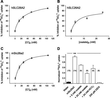
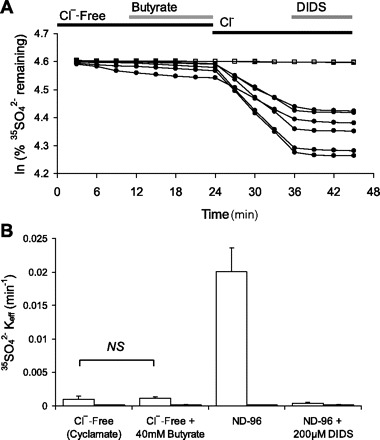
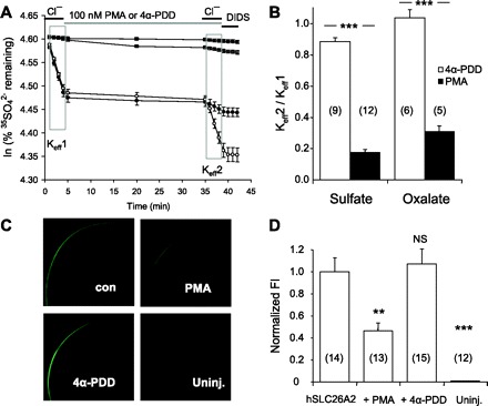
Nephrolithiasis in the Slc26a6(-/-) mouse is accompanied by 50-75% reduction in intestinal oxalate secretion with unchanged intestinal oxalate absorption. The molecular identities of enterocyte pathways for oxalate absorption and for Slc26a6-independent oxalate secretion remain undefined. The reported intestinal expression of SO(4)(2-) transporter SLC26A2 prompted us to characterize transport of oxalate and other anions by human SLC26A2 and mouse Slc26a2 expressed in Xenopus oocytes. We found that hSLC26A2-mediated [(14)C]oxalate uptake (K(1/2) of 0.65 +/- 0.08 mM) was cis-inhibited by external SO(4)(2-) (K(1/2) of 3.1 mM). hSLC26A2-mediated bidirectional oxalate/SO(4)(2-) exchange exhibited extracellular SO(4)(2-) K(1/2) of 1.58 +/- 0.44 mM for exchange with intracellular [(14)C]oxalate, and extracellular oxalate K(1/2) of 0.14 +/- 0.11 mM for exchange with intracellular (35)SO(4)(2-). Influx rates and K(1/2) values for mSlc26a2 were similar. hSLC26A2-mediated oxalate/Cl(-) exchange and bidirectional SO(4)(2-)/Cl(-) exchange were not detectably electrogenic. Both SLC26A2 orthologs exhibited nonsaturable extracellular Cl(-) dependence for efflux of intracellular [(14)C]oxalate, (35)SO(4)(2-), or (36)Cl(-). Rate constants for (36)Cl(-) efflux into extracellular Cl(-), SO(4)(2-), and oxalate were uniformly 10-fold lower than for oppositely directed exchange. Acidic extracellular pH (pH(o)) inhibited all modes of hSLC26A2-mediated anion exchange. In contrast, acidic intracellular pH (pH(i)) selectively activated exchange of extracellular Cl(-) for intracellular (35)SO(4)(2-) but not for intracellular (36)Cl(-) or [(14)C]oxalate. Protein kinase C inhibited hSLC26A2 by reducing its surface abundance. Diastrophic dysplasia mutants R279W and A386V of hSLC26A2 exhibited similar reductions in uptake of both (35)SO(4)(2-) and [(14)C]oxalate. A386V surface abundance was reduced, but R279W surface abundance was at wild-type levels.


Figures

Fig. 1.
Fig. 1.
Cis-inhibition of SLC26A2-mediated SO42− uptake by extracellular anions. A: extracellular [Cl] dependence for cis-inhibition of 35SO42− uptake by hSLC26A2. K1/2 = 24.1 ± 3.8 mM Cl (R2 = 0.90, n = 12–14). B: extracellular [oxalate] dependence for cis-inhibition of 35SO42− uptake by hSLC26A2. K1/2 = 5.1 ± 1.7 mM oxalate (R2 = 0.86, n = 7–9). C: extracellular [Cl] dependence for cis-inhibition of 35SO42− uptake by mSlc26a2. K1/2 = 18.4 ± 3.4 mM Cl (R2 = 0.93, n = 6–7). D: cis-inhibition of mSlc26a2-mediated 35SO42− uptake by oxalate or HCO3 at the indicated concentrations for (n) oocytes compared with complete inhibition by DIDS (200 μM). Oocytes previously injected with 50 ng cRNA or water were subjected to 30 min uptake in the presence of 1 mM extracellular SO42−. Uptake data were normalized to same-day experiments carried out in cyclamate. Solid bar indicates water-injected oocytes. Normalized uninhibited SO42− uptake was (in nmol/oocyte−1·h−1) 1.42 ± 0.05 (A), 1.48 ± 0.07 (B), 1.13 ± 0.05 (C), and 1.03 ± 0.19 (D). Values are means ± SE. **P < 0.01.
Fig. 2.
Fig. 2.
hSLC26A2 mediates bidirectional oxalate/SO42− exchange. A: bath [oxalate] dependence of 30 min oxalate uptake by oocytes expressing hSLC26A2. K1/2 = 0.65 ± 0.08 mM oxalate, Hill coefficient = 2.01 ± 0.41 (R2 = 0.66, n = 19–20). Normalized oxalate uptake = 1.0 represented 0.15 nmol/h. B: bath [SO42−] dependence for cis-inhibition of 1 mM bath oxalate uptake by oocytes expressing hSLC26A2. K1/2 = 3.07 ± 0.74 mM sulfate (R2 = 0.72, n = 7–17). C: traces of 35SO42− efflux from individual oocytes previously injected with water (open squares) or hSLC26A2 (filled circles) during sequential exposure to Cl-free cyclamate baths in the initial presence and subsequent absence of 25 mM oxalate. D: traces of [14C]oxalate efflux from individual oocytes previously injected with water (open squares) or hSLC26A2 (filled circles) during sequential exposure to Cl-free baths in the initial presence and subsequent absence of 64 mM SO42−. Oocytes previously injected with 50 ng cRNA or water were assayed 4 days postinjection. Values are means ± SE.
Fig. 3.
Fig. 3.
[Substrate] dependence of SLC26A2-mediated oxalate/SO42− exchange. A: bath [oxalate] dependence of hSLC26A2-mediated 35SO42− efflux. K1/2 = 1.58 ± 0.44 mM oxalate (n = 4–5). B: bath [SO42−] dependence of hSLC26A2-mediated oxalate efflux. K1/2 = 0.14 ± 0.05 mM SO42− (n = 5–22). C: bath [oxalate] dependence of mSlc26a2-mediated 35SO42− efflux. K1/2 = 3.74 ± 0.73 mM oxalate (n = 3–15). D: bath [SO42−] dependence of mSlc26a2-mediated oxalate efflux. K1/2 = 0.20 ± 0.08 mM SO42− (n = 4–20). Oocytes previously injected with 50 ng cRNA were assayed 3–4 days postinjection. Values are means ± SE. Curve fits in B and D were unchanged by inclusion of additional [14C]oxalate efflux rate constants measured at higher bath [SO42−] values of 8 and 9 mM (n = 5–6, data not shown). There was no sulfate self-inhibition at still higher bath [SO42−] values of 16, 32, or 64 mM (not shown).
Fig. 4.
Fig. 4.
SLC26A2-mediated exchange of intracellular 35SO42− for bath Cl exhibits very low Cl affinity. A: traces of 35SO42− efflux from individual oocytes previously injected with water (open squares, n = 2) or hSLC26A2 cRNA (filled circles, n = 6) during sequential increases in bath [Cl], followed by exposure to 200 μM DIDS. B: extracellular Cl concentration ([Cl]o) dependence of hSLC26A2-mediated [35SO42−]i/[Cl]o exchange determined from A. C: traces of 35SO42− efflux from individual oocytes previously injected with water (open squares, n = 2) or mSlc26a2 cRNA (filled circles, n = 6) during sequential increases in bath [Cl], followed by addition of 200 μM DIDS. D: [Cl]o dependence of mSlc26a2-mediated [35SO42−]i/[Cl]o exchange determined from C. Values in B and D are means ± SE measured 3 days after oocyte injection with cRNA encoding hSLC26A2 (1 ng) or mSlc26a2 (5 ng). Data are representative of 2–3 similar experiments.
Fig. 5.
Fig. 5.
hSLC26A2-mediated exchange of intracellular [14C]oxalate or 36Cl for bath Cl exhibits low bath Cl affinity. A: traces of [14C]oxalate efflux from individual oocytes previously injected with water (open squares, n = 3) or hSLC26A2 cRNA (closed circles, n = 6) during sequential increases in bath [Cl], followed by addition of 200 μM DIDS. B: [Cl]o dependence of hSLC26A2-mediated [14C]oxalatei/Clo exchange determined from A. C: traces of 36Cl efflux from individual oocytes previously injected with water (open squares, n = 3) or hSLC26A2 cRNA (filled circles, n = 6) during sequential increases in bath [Cl], followed by exposure to 200 μM DIDS. D: [Cl]o dependence of hSLC26A2-mediated 36Cli/Cloexchange determined from C. Values in B and D are means ± SE measured 3 days after oocyte injection with 1 ng hSLC26A2 cRNA and are representative of 2–3 similar experiments.
Fig. 6.
Fig. 6.
SLC26A2 mediates electroneutral exchange of bath oxalate or bath sulfate for nominal intracellular Cl (Cli). A: steady-state current-voltage (I-V) relationships of hSLC26A2-expressing oocytes measured first in Na gluconate bath (open circles) and again after subsequent bath addition of 5 mM oxalate (filled circles; n = 5). B: steady-state I-V relationships of hSLC26A2-expressing oocytes during sequential exposure to baths containing Na cyclamate (open circles), cyclamate with 5 mM SO42− (filled circles), and 64 mM SO42− (open triangles), with subsequent addition of 200 μM DIDS (filled triangles; n = 10). C: steady-state I-V relationships of mSlc26a2-expressing oocytes measured first in Na gluconate bath (open circles) and again after subsequent bath addition of 5 mM oxalate (filled circles; n = 4). D: steady-state I-V relationships of mSlc26a2-expressing oocytes during sequential exposure to baths containing Na cyclamate (open circles), cyclamate with 5 mM SO42− (filled circles), and 64 mM SO42− (open triangles), with subsequent addition of 200 μM DIDS (filled triangles; n = 9). Values are means ± SE.
Fig. 7.
Fig. 7.
Regulation of hSLC26A2-mediated anion exchange by extracellular pH. A: 35SO42− efflux traces from individual oocytes previously injected with water (open squares, n = 3) or with 0.5 ng hSLC26A2 (filled circles, n = 5) during sequential exposure to ND96 bath at pH 5.0, pH 8.0, and subsequent addition of 200 μM DIDS. B: 35SO42− efflux rate constants of oocytes expressing hSLC26A2 during sequential exposure to ND96 baths of pH 5.0, pH 8.0, and after addition of DIDS (open bars, n = 16). Solid bars show water-injected oocytes (n = 5). These data include those of A. C: 36Cl efflux rate constants of oocytes expressing hSLC26A2 (10 ng cRNA) during sequential exposure to ND96 baths of pH 5.0, pH 8.0, and after addition of DIDS (open bars, n = 12). Solid bars show water-injected oocytes (n = 6). D: [14C]oxalate efflux rate constants of oocytes expressing hSLC26A2 (50 ng cRNA) during sequential exposure to ND96 baths of pH 5.0, pH 8.0, and after addition of DIDS (open bars, n = 11). Solid bars show water-injected oocytes (n = 4). Values in BD are means ± SE measured 3–4 days post-cRNA injection.
Fig. 8.
Fig. 8.
Regulation of hSLC26A2-mediated anion exchange by intracellular pH. A: 35SO42− efflux traces from individual uninjected oocytes (open squares, n = 2) or from oocytes previously injected with 0.5 ng hSLC26A2 cRNA (closed circles, n = 4) during sequential exposure to baths containing 64 mM Cl with 40 mM Na butyrate, 64 mM Cl with 40 mM sodium cyclamate, and Cl-free cyclamate. B: effect of butyrate removal (intracellular alkalinization) on 35SO42−i/Clo exchange by oocytes previously injected with 0.5 ng hSLC26A2 (open bars, n = 10) or from uninjected oocytes (solid bars, n = 2) determined as in A. C: effect of butyrate removal on Cli/Clo exchange by oocytes previously injected with 10 ng hSLC26A2 (open bars, n = 11) and by uninjected (solid bars, n = 5). D: effect of butyrate removal on [14C]oxalatei/Clo exchange from oocytes previously injected with 50 ng hSLC26A2 (open bars, n = 11) and by uninjected oocytes (solid bars, n = 6). Values in BD are means ± SE measured 3–4 days post-cRNA injection. All oocytes were preincubated 30 min in Cl-free 40 mM Na butyrate before initiation of efflux experiments.
Fig. 9.
Fig. 9.
hSLC26A2 does not mediate 35SO42−i/butyrateo exchange. A. 35SO42− efflux traces from individual uninjected oocytes (open squares, n = 2) or from oocytes previously injected with 0.5 ng hSLC26A2 cRNA (closed circles, n = 7) during sequential exposure to baths containing Cl-free cyclamate, cyclamate with 40 mM Na butyrate, butyrate-free ND96, and ND96 containing 200 μM DIDS. B: 35SO42− efflux rate constants (means ± SE) of oocytes shown in A. Values are measured 3 days post-cRNA injection.
Fig. 10.
Fig. 10.
Protein kinase C inhibits hSLC26A2-mediated efflux of SO42− and of oxalate by reducing surface expression. A: 35SO42− efflux traces averaged from representative individual uninjected oocytes (two top traces, each n = 3) or from oocytes previously injected with 0.5 ng hSLC26A2 cRNA (two bottom traces, each n = 6) during sequential exposure to baths containing ND96, Cl-free cyclamate containing PMA (closed symbols) or 4α-phorbol-didecanoate (4α-PDD) (open symbols), then ND96 in the continued presence of PMA or 4α-PDD, and finally, ND96 with 200 μM DIDS. Efflux trace slopes from the first and second bath Cl periods (boxed areas) yield Keff1 and Keff2 as indicated. B: ratios of mean rate constants for 35SO42− efflux (Keff2/Keff1) into ND96 in the presence of 4α-PDD (open bars) or PMA (solid bars). C: confocal fluorescence micrographs of representative oocytes, uninjected or previously injected with 10 ng hSLC26A2-GFP cRNA, then incubated 30 min in the absence (con) or presence of PMA or 4α-PDD as indicated. D: normalized hSLC26A6-GFP fluorescence intensity (FI, means ± SE) at the periphery of (n) oocytes treated with PMA or 4α-PDD. Untreated hSLC26A2-GFP-expressing oocytes were assigned a mean FI value of 1.0. **P < 0.01; ***P < 0.001.
Fig. 11.
Fig. 11.
Two diastrophic dysplasia sulfate transporter (DTDST) mutants of hSLC26A2 exhibit impaired efflux of 35SO42− and of oxalate but differ in surface expression. A: normalized 35SO42− uptake by (n) uninjected oocytes or oocytes expressing wild-type (WT) hSLC26A2 or the indicated hSLC26A2 mutants (0.5 ng cRNA). WT uptake 0.17 ± 0.02 nmol/h. B: normalized [14C]oxalate uptake by (n) uninjected oocytes or oocytes expressing WT or mutant hSLC26A2 (50 ng cRNA). WT uptake was 0.20 ± 0.02 nmol/h. C: confocal immunofluorescence micrographs of representative oocytes expressing WT hSLC26A2 or the indicated mutants. D: normalized mean fluorescence intensity (FI) measured at the periphery of (n) oocytes expressing WT or mutant SLC26A2. WT hSLC26A2 intensity was assigned a mean FI of 1.0. ***P < 0.001.

Similar articles

Cited by

References

    1. Aronson PS, Giebisch G. Mechanisms of chloride transport in the proximal tubule. Am J Physiol Renal Physiol 273: F179–F192, 1997 - PubMed
    1. Barmeyer C, Ye JH, Sidani S, Geibel J, Binder HJ, Rajendran VM. Characteristics of rat downregulated in adenoma (rDRA) expressed in HEK 293 cells. Pflügers Arch 454: 441–450, 2007 - PubMed
    1. Beauge L, DiPolo R. The squid axon Na/Ca2+ exchanger shows ping pong kinetics only when the Ca2+(i)-regulatory site is saturated. Cell Physiol Biochem 23: 37–42, 2009 - PubMed
    1. Burckhardt BC, Fromter E. Pathways of NH3/NH4+ permeation across Xenopus laevis oocyte cell membrane. Pflügers Arch 420: 83–86, 1992 - PubMed
    1. Chapman JM, Karniski LP. Protein localization of SLC26A2 (DTDST) in rat kidneys (Abstract). FASEB J 21: 937. 27, 2007 - PubMed

Publication types

MeSH terms