. 2010 May 1;21(9):1494-504.
doi: 10.1091/mbc.e09-12-1023. Epub 2010 Mar 10.

Stepwise assembly of dimeric F(1)F(o)-ATP synthase in mitochondria involves the small F(o)-subunits k and i

Affiliations

Stepwise assembly of dimeric F(1)F(o)-ATP synthase in mitochondria involves the small F(o)-subunits k and i

Karina Wagner et al. Mol Biol Cell. .

Abstract

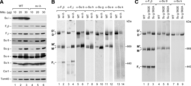
F(1)F(o)-ATP synthase is a key enzyme of oxidative phosphorylation that is localized in the inner membrane of mitochondria. It uses the energy stored in the proton gradient across the inner mitochondrial membrane to catalyze the synthesis of ATP from ADP and phosphate. Dimeric and higher oligomeric forms of ATP synthase have been observed in mitochondria from various organisms. Oligomerization of ATP synthase is critical for the morphology of the inner mitochondrial membrane because it supports the generation of tubular cristae membrane domains. Association of individual F(1)F(o)-ATP synthase complexes is mediated by the membrane-embedded F(o)-part. Several subunits were mapped to monomer-monomer-interfaces of yeast ATP synthase complexes, but only Su e (Atp21) and Su g (Atp20) have so far been identified as crucial for the formation of stable dimers. We show that two other small F(o)-components, Su k (Atp19) and Su i (Atp18) are involved in the stepwise assembly of F(1)F(o)-ATP synthase dimers and oligomers. We have identified an intermediate form of the ATP synthase dimer, which accumulates in the absence of Su i. Moreover, our data indicate that Su i facilitates the incorporation of newly synthesized subunits into ATP synthase complexes.


Figures

Figure 1.
Figure 1.
Su k differentially associates with different forms of F1Fo-ATP synthase. (A) Mitochondria isolated from a wild-type (WT) S. cerevisiae strain were solubilized in 1% digitonin, protein complexes were separated on a 4–8% polyacrylamide blue native gel and analyzed by immunoblotting with the indicated antisera. (B) 35S-Labeled Su k was imported into isolated wild-type (WT), su gΔ, and su eΔ mitochondria in the presence or absence of a Δψ. On treatment with proteinase K mitochondria were solubilized in digitonin-containing buffer and analyzed by BN-PAGE and digital autoradiography. (C) WT, su eΔ, and su gΔ mutant mitochondria were analyzed by BN-PAGE as described in A. (D) Mitochondria were incubated with radiolabeled Su k in the presence or absence of Δψ. After treatment with proteinase K, samples were analyzed by SDS-PAGE and digital autoradiography. (E) Mitochondria were treated with 0.1 M sodium carbonate, pH 10.8. Membrane-integral and soluble proteins were separated by ultracentrifugation (125,000 × g). Total (T), pellet (P), and supernatant (S) fractions were analyzed by SDS-PAGE and Western blotting with the indicated antibodies. M, monomeric F1Fo-ATP synthase; M', primed monomer; D' and D, dimeric forms of F1Fo-ATP synthase.
Figure 2.
Figure 2.
Su k is required for the formation of stable ATP synthase dimers. (A) Analysis of steady-state protein levels of wild-type (WT) and su kΔ mitochondria. Ten, 20, and 30 μg of mitochondrial proteins were analyzed by SDS-PAGE and Western blotting. (B) Isolated wild-type (WT) and su kΔ mitochondria were solubilized in buffer containing 1% digitonin and analyzed by BN-PAGE and immunoblotting with the indicated antisera. M, monomeric F1Fo-ATP synthase; M', primed monomer; D' and D, dimeric forms of F1Fo-ATP synthase.
Figure 3.
Figure 3.
The primed monomer of F1Fo-ATP synthase accumulates in the absence of Su k. (A) Import of radiolabeled F1γ into wild-type and su kΔ mitochondria in the presence or absence of a Δψ. After import reactions mitochondria were treated with proteinase K and solubilized in digitonin buffer. Protein extracts were separated on 4–8% polyacrylamide blue native gels and analyzed by digital autoradiography. (B–D) Import of Su h, Su e, and Su g was performed and analyzed as described in A. Note that the presence of Δψ is not essential for import and assembly of Su e (Wagner et al., 2009). M, monomeric F1Fo-ATP synthase; M', primed monomer; D' and D, dimeric forms of F1Fo-ATP synthase; F1, free F1-subcomplex.
Figure 4.
Figure 4.
Higher oligomeric states of ATP synthase depend on Su k. (A) Mitochondria were solubilized in 0.35% digitonin and subjected to BN-PAGE by using 3–13% polyacrylamide gels. Activity staining of F1Fo-ATP synthase complexes (lane 1) was carried out as described in Materials and Methods. 35S-Labeled Su h was imported into wild-type (WT) and su kΔ mitochondria in the presence or absence of a Δψ. After solubilization of mitochondria in digitonin buffer (0.35%) and BN-PAGE, gels were analyzed by digital autoradiography. (B) Import of radiolabeled Su h into WT, su gΔ, and su eΔ mitochondria was performed and analyzed as outlined in A. M, ATP synthase monomers; D, ATP synthase dimers; O, oligomeric forms of ATP synthase; F1, F1-subcomplex; arrowhead, partially fragmented oligomer. The gel system used here to detect larger oligomeric forms does not clearly separate the distinct forms of ATP synthase monomers and dimers.
Figure 5.
Figure 5.
Su i is required for the formation of a distinct F1Fo-ATP synthase dimer form. (A) Analysis of steady-state protein levels in wild-type (WT) and su iΔ mitochondria. Ten, 20, and 30 μg of mitochondrial proteins were analyzed by SDS-PAGE and immunoblotting. (B) Digitonin-solubilized wild-type (WT) and su iΔ mitochondria were subjected to high-resolution BN-PAGE and analyzed by Western blotting with the indicated antisera. (C) Isolated WT, Su g S62E, and Su g S62A mitochondria were solubilized in buffer containing 1% digitonin. Protein complexes were separated on 4–8% polyacrylamide blue native gels and analyzed by immunoblotting with antisera against F1β, Su 9 and Su k. M, monomeric ATP synthase; M', primed monomer; D' and D, dimeric forms of F1Fo-ATP synthase; F1, detached F1-subcomplex.
Figure 6.
Figure 6.
Independent assembly of Su i and Su e/g with monomeric ATP synthase. (A) Wild-type (WT), su gΔ, and su eΔ mitochondria were incubated with 35S-labeled Su i in the presence or absence of a Δψ. Subsequent to proteinase K treatment, mitochondria were solubilized in digitonin-containing buffer. Samples were applied to a 4–8% blue native gel and protein complexes were visualized by digital autoradiography. (B and C) Radiolabeled Su e and Su g were imported into WT and su iΔ mitochondria. Samples were analyzed as described in A. M, monomeric ATP synthase; M', primed monomer; D' and D, dimeric forms of F1Fo-ATP synthase.
Figure 7.
Figure 7.
Su i facilitates the incorporation of imported subunits into central and peripheral stalk regions. 35S-Labeled F1γ (A), F1δ (B), and Su h (C) were imported into wild-type (WT) and su iΔ mitochondria in the presence or absence of a Δψ. On treatment with proteinase K and solubilization of mitochondria in digitonin buffer, samples were subjected to BN-PAGE (A–C, top) or SDS-PAGE (A–C, bottom). Visualization of imported proteins was done by digital autoradiography. M, monomeric ATP synthase; M', primed monomer; D' and D, dimeric forms of F1Fo-ATP synthase; F1, F1-subcomplex.
Figure 8.
Figure 8.
Su i is required for the assembly of newly synthesized Su k and Su b with F1Fo-ATP synthase. (A) Wild-type (WT) and su iΔ mutant mitochondria were incubated with the radiolabeled precursor of Su k in the presence or absence of a Δψ. After proteinase K-treatment, mitochondria were either solubilized in digitonin-containing buffer and analyzed by BN-PAGE (top) or denatured for analysis by SDS-PAGE (bottom). Digital autoradiography was used for the detection of imported proteins. (B) Import of 35S-labeled Su b was carried out and analyzed as described in A. (C) 35S-Labeled Su b was imported into WT, su gΔ, and su eΔ mitochondria and analyzed by BN-PAGE and digital autoradiography as described in A. M, monomeric F1Fo-ATP synthase; M', primed monomer; D' and D, dimeric forms of ATP synthase.
Figure 9.
Figure 9.
Model for stepwise assembly of dimeric F1Fo-ATP synthase in mitochondria. Sequential association of Su e and Su g with monomeric ATP synthase (M) leads to the formation of a primed monomer (M'). An intermediate dimeric form (D') assembles from these primed monomers. Su i drives the conversion of the intermediate dimer to the mature dimer (D), which stably associates with Su k. In addition to its role in dimer maturation, Su i facilitates the incorporation of new subunits into F1Fo-ATP synthase already at the level of the monomeric form. Beige, inner mitochondrial membrane; blue, F1-part of ATP synthase; light green, Fo-part of ATP synthase with peripheral stalk; dark green, Su i at the monomeric ATP synthase facilitating subunit exchange; red, Su e and Su g; yellow, Su i mediating the transition from D' to D; and orange, Su k.

References

    1. Acín-Pérez R., Fernández-Silva P., Peleato M. L., Pérez-Martos A., Enriquez J. A. Respiratory active mitochondrial supercomplexes. Mol. Cell. 2008;32:529–539. - PubMed
    1. Allen R. D., Schroeder C. C., Fok A. K. An investigation of mitochondrial inner membranes by rapid-freeze deep-etch techniques. J. Cell Biol. 1989;108:2233–2240. - PMC - PubMed
    1. Allen R. D. Membrane tubulation and proton pumps. Protoplasma. 1995;189:1–8.
    1. Amutha B., Gordon D. M., Gu Y., Pain D. A novel role of Mgm1p, a dynamin-related GTPase, in ATP synthase assembly and cristae formation/maintenance. Biochem. J. 2004;381:19–23. - PMC - PubMed
    1. Arnold I., Pfeiffer K., Neupert W., Stuart R. A., Schägger H. Yeast mitochondrial F1F0-ATP synthase exists as a dimer: identification of three dimer-specific subunits. EMBO J. 1998;17:7170–7178. - PMC - PubMed

Publication types

MeSH terms

Substances